

 PDF
PDF ePub
ePub Citation
 Print
Print


Abstract
A 77-year-old woman presented with bilateral leg weakness, accompanied by severe axial back and radicular pain, after a L4–5 epidural injection. She had been receiving misappropriated epidural injections for the last few months. A contrast-enhanced magnetic resonance image showed rim enhancing, spinal canal compromising cystic lesion at the posterior epidural space of L4–5. During surgery, a severely central compromised non-communicating cystic lesion located at posterior epidural space was resected. A histological report of this lesion confirmed a pseudocyst containing a degenerated synovial tissue. Herein, we report our experience of cauda equine syndrome after epidural injection with successful treatment.
Figures and Tables
Figure 1
The plane lateral radiograph (A) and sagittal T2WI magnetic resonance imaging (B) demonstrate L4 degenerative spondylolisthesis and central canal stenosis at L4–5.
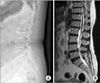
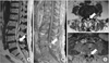
Figure 2
Contrast magnetic resonance imaging demonstrates a posterior epidural cystic lesion at L4–5. Sagittal T1WI shows a low signal (A) and T2WI shows a high signal intensity of the mass (arrows) (B), suggesting the facet synovial cyst. This intraspinal cystic lesion was located on the posterior epidural space (arrows) on axial T1WI (C) and T2WI (D).
References
1. Baker JK, Hanson GW. Cyst of the ligamentum flavum. Spine (Phila Pa 1976). 1994; 19:1092–1094.

2. Doyle AJ, Merrilees M. Synovial cysts of the lumbar facet joints in a symptomatic population: prevalence on magnetic resonance imaging. Spine (Phila Pa 1976). 2004; 29:874–878.
3. Asamoto S, Jimbo H, Fukui Y, et al. Cyst of the ligamentum flavum: case report. Neurol Med Chir (Tokyo). 2005; 45:653–656.
4. Farfan HF, Sullivan JD. The relation of facet orientation to intervertebral disc failure. Can J Surg. 1967; 10:179–185.
5. Kirkaldy-Willis WH, Wedge JH, Yong-Hing K, Reilly J. Pathology and pathogenesis of lumbar spondylosis and stenosis. Spine (Phila Pa 1976). 1978; 3:319–328.
6. Howington JU, Connolly ES, Voorhies RM. Intraspinal synovial cysts: 10-year experience at the Ochsner Clinic. J Neurosurg. 1999; 91:S193–S199.
7. Okada A, Harata S, Takeda Y, Nakamura T, Takagaki K, Endo M. Age-related changes in proteoglycans of human ligamentum flavum. Spine (Phila Pa 1976). 1993; 18:2261–2266.
8. Haase J. Extradural cyst of ligamentum flavum L 4: a case. Acta Orthop Scand. 1972; 43:32–38.
9. Métellus P, Fuentes S, Adetchessi T, et al. Retrospective study of 77 patients harbouring lumbar synovial cysts: functional and neurological outcome. Acta Neurochir (Wien). 2006; 148:47–54. discussion 54.
10. Manchikanti L, Abdi S, Atluri S, et al. An update of comprehensive evidence-based guidelines for interventional techniques in chronic spinal pain. Part II: guidance and recommendations. Pain Physician. 2013; 16:S49–283.
 XML Download