

 PDF
PDF ePub
ePub Citation
 Print
Print


Abstract
Purpose
Soft tissue reconstruction of a defect around the foot and ankle is a particularly challenging procedure due to the anatomical and functional characteristics of this area. Hence, only a limited number of treatment options are available. Moreover, if patients wish to avoid additional scars on the ipsilateral lower leg for cosmetic reasons, even fewer options are available for treatment. The authors used an anterolateral thigh perforator flap for soft tissue defects in this area, when other surgical options were inadequate. The aim of this study was to report the clinical results and the efficacy of this procedure.
Materials and Methods
Sixteen cases of soft tissue defects around the foot and ankle were included. Participants included 12 male and 4 female subjects, and the mean age was 34 years. The most common cause of defect was acute trauma, and the average follow-up period was 33 months. Flap survival time, surgical complications, and ambulation status at the final follow-up stage were evaluated.
Results
All 16 flaps successfully survived, except for one case with partial flap necrosis that was thought to be due to weight bearing earlier than scheduled. All patients were able to walk independently without any aid at the final follow-up stage. No patients showed other significant surgical complications.
Figures and Tables
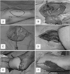 | Figure 1(A) The flap size was designed to be 16×19 cm in an elliptical shape. (B) The flap was dissected from the donor site. The descending branch of the lateral circumflex femoral artery and perforator artery was seen between the flap and the donor site. (C) The flap was detached from the donor site. The perforator artery was seen on the bottom of flap attachment. (D, E) The end-to-end anastomosis of 2 veins and 1 artery was done between the recipient site and the flap. (F) Splitthickness skin graft was done on the donor site.
|
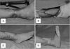 | Figure 2(A) A 8×5 cm sized soft tissue defect on the anterior ankle. (B) Anterolateral thigh flap application. (C, D) Twenty months after the anterolateral thigh flap surgery with fully recovered range of motion.
|
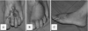 | Figure 3(A) Scar contracture on the dorsum of the left foot. (B, C) Seven months after the anterolateral thigh flap surgery.
|
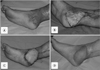 | Figure 4(A) Chronic inflammatory reaction with superficial necrosis on the posterior ankle. (B) Debridement of necrotic tissue. A 15×7 cm sized anterolateral thigh flap (C), 12 months after the surgery (D).
|
References
1. Jeng SF, Wei FC, Kuo YR. Salvage of the distal foot using the distally based sural island flap. Ann Plast Surg. 1999; 43:499–505.

2. Lee JM, Kim MK. Reconstruction of the extremities with the dorsalis pedis free flap. J Korean Microsurg Soc. 1999; 8:77–83.
3. Pontén B. The fasciocutaneous flap: its use in soft tissue defects of the lower leg. Br J Plast Surg. 1981; 34:215–220.
4. Donski PK, Fogdestam I. Distally based fasciocutaneous flap from the sural region. A preliminary report. Scand J Plast Reconstr Surg. 1983; 17:191–196.
5. Masquelet AC, Beveridge J, Romana C, Gerber C. The lateral supramalleolar flap. Plast Reconstr Surg. 1988; 81:74–81.
6. Yamada N, Kakibuchi M, Kitayoshi H, Matsuda K, Yano K, Hosokawa K. A new way of elevating the anterolateral thigh flap. Plast Reconstr Surg. 2001; 108:1677–1682.
7. Song YG, Chen GZ, Song YL. The free thigh flap: a new free flap concept based on the septocutaneous artery. Br J Plast Surg. 1984; 37:149–159.
8. Koshima I, Moriguchi T, Fukuda H, Yoshikawa Y, Soeda S. Free, thinned, paraumbilical perforator-based flaps. J Reconstr Microsurg. 1991; 7:313–316.
9. Soeda S. Practical guide for the tubed pedicle flap reconstruction. Keisei Geka. 1970; 13:457–463.
10. Santanelli F, Tenna S, Pace A, Scuderi N. Free flap reconstruction of the sole of the foot with or without sensory nerve coaptation. Plast Reconstr Surg. 2002; 109:2314–2322. discussion 2323-4.
11. Mahoney J. Complications of free flap donor sites. Microsurgery. 1995; 16:437–444.
12. Lee KS, Wie DG, Han SW. Morbidity of the foot as a free-flap donor site. J Korean Microsurg Soc. 1997; 6:39–46.
13. Mardini S, Wei FC, Salgado CJ, Chen HC. Reconstruction of the reconstructive ladder. Plast Reconstr Surg. 2005; 115:2174.
14. Kwon BK, Chung DW, Lee JH, Choi IH, Song JH, Lee SW. One-stage reverse lateral supramalleolar adipofascial flap for soft tissue reconstruction of the foot and ankle joint. J Korean Microsurg Soc. 2007; 16:93–99.
15. Hallock GG. The mangled foot and ankle: soft tissue salvage techniques. Clin Podiatr Med Surg. 2014; 31:565–576.
16. Kermarrec G, Masquelet AC. Sural flap for coverage of a softtissue defect of a leg with an occluded fibular artery: a case report. J Plast Reconstr Aesthet Surg. 2014; 67:729–731.
17. Shanahan RE, Gingrass RP. Medial plantar sensory flap for coverage of heel defects. Plast Reconstr Surg. 1979; 64:295–298.
18. Zhu YL, Wang Y, He XQ, Zhu M, Li FB, Xu YQ. Foot and ankle reconstruction: an experience on the use of 14 different flaps in 226 cases. Microsurgery. 2013; 33:600–604.
19. Kong BS, Seo MS, Ha JM. Thigh perforator free flap for reconstruction of the soft tissue defect of the lower extremity. J Korean Foot Ankle Soc. 2007; 11:232–237.
20. Koshima I, Fukuda H, Utunomiya R, Soeda S. The anterolateral thigh flap; variations in its vascular pedicle. Br J Plast Surg. 1989; 42:260–262.
21. Collins J, Ayeni O, Thoma A. A systematic review of anterolateral thigh flap donor site morbidity. Can J Plast Surg. 2012; 20:17–23.
22. Kimata Y, Uchiyama K, Ebihara S, Nakatsuka T, Harii K. Anatomic variations and technical problems of the anterolateral thigh flap: a report of 74 cases. Plast Reconstr Surg. 1998; 102:1517–1523.
23. Wei FC, Jain V, Celik N, Chen HC, Chuang DC, Lin CH. Have we found an ideal soft-tissue flap? An experience with 672 anterolateral thigh flaps. Plast Reconstr Surg. 2002; 109:2219–2226. discussion 2227-30.
24. Kuo YR, Seng-Feng J, Kuo FM, Liu YT, Lai PW. Versatility of the free anterolateral thigh flap for reconstruction of soft-tissue defects: review of 140 cases. Ann Plast Surg. 2002; 48:161–166.
25. Yildirim S, Avci G, Aköz T. Soft-tissue reconstruction using a free anterolateral thigh flap: experience with 28 patients. Ann Plast Surg. 2003; 51:37–44.
26. Kim HS, Kim KC, Kim SE. Soft tissue reconstruction of children's extremity with perforator free flap. J Korean Microsurg Soc. 2007; 16:14–22.
 XML Download