Abstract
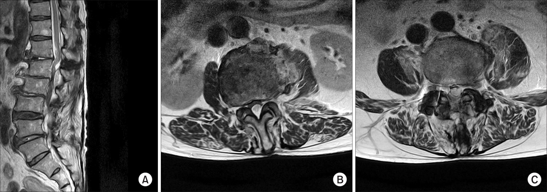
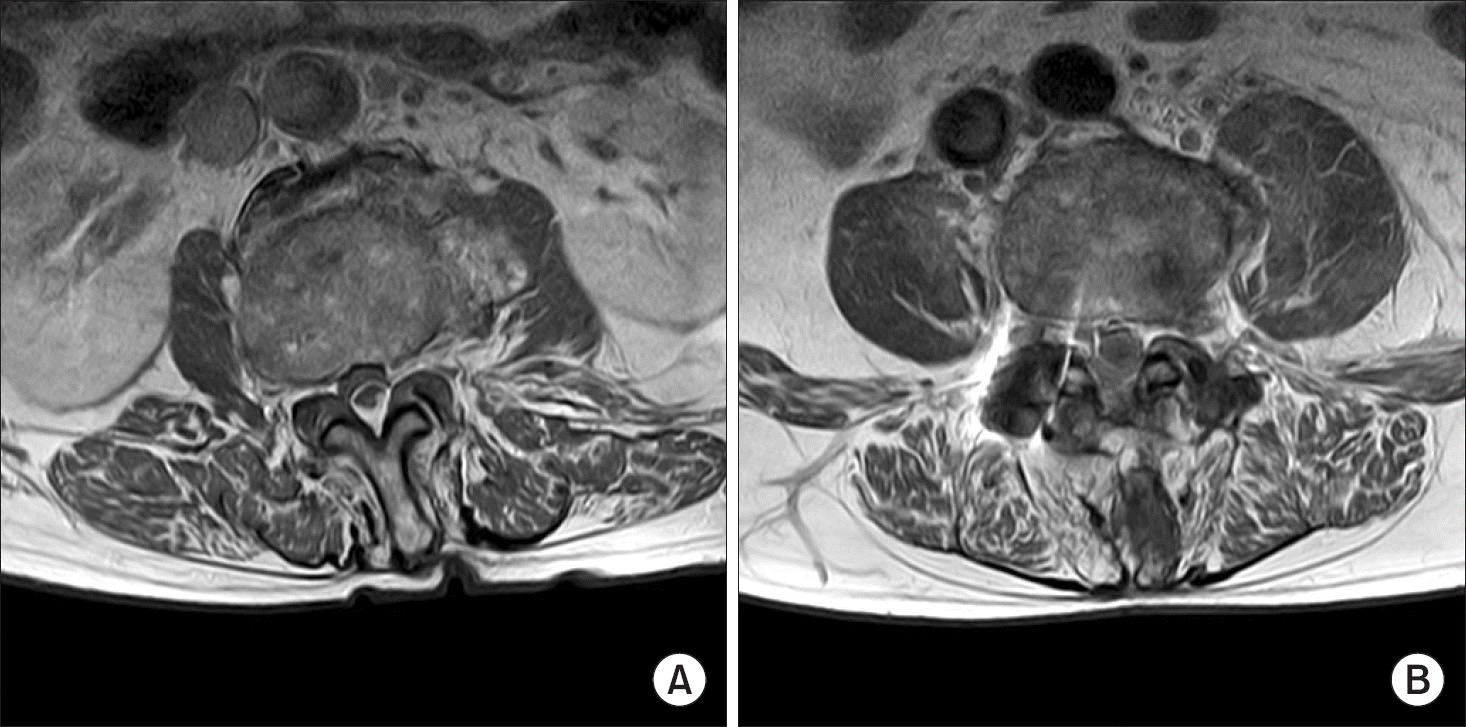
Spinal infection due to Serratia marcescens is very rare. A 78-year-old male patient withoutany risk factor was admitted to our hospital with chief complaints of severe back pain, fever, weakness in both legs, and bowel dysfunction, following caudal epidural injection. Magnetic resonance imaging revealed spondylodiscitis with epidural abscess. Surgical decompression was performed and the epidural abscess was removed. The cultures isolated S. marcescens, which can cause nosocomial infection in immunocompromised patient. However, to the best of our knowledge, we report the first case of S. marcescens spinal epidural abscess following epidural injection, with literature review.
Go to : 
REFERENCES
2. Rigamonti D, Liem L, Sampath P. . Spinal epidural abscess: contemporary trends in etiology, evaluation, and management. Surg Neurol. 1999; 52:189–96.

4. Reihsaus E, Waldbaur H, Seeling W. Spinal epidural abscess: a meta-analysis of 915 patients. Neurosurg Rev. 2000; 23:175–204. discussion 205.

5. Soehle M, Wallenfang T. Spinal epidural abscesses: clinical manifestations, prognostic factors, and outcomes. Neurosurgery. 2002; 51:79–85. discussion 86, 7.

6. Hadjipavlou AG, Gaitanis IN, Papadopoulos CA, Katonis PG, Kontakis GM. Serratia spondylodiscitis after elective lumbar spine surgery: a report of two cases. Spine (Phila Pa 1976). 2002; 27:E507–12.
7. Alfonso Olmos M, Silva González A, Duart Clemente J, Villas Tomé C. Infected vertebroplasty due to uncommon bacteria solved surgically: a rare and threatening life complication of a common procedure: report of a case and a review of the literature. Spine (Phila Pa 1976). 2006; 31:E770–3.
8. Weinstein MA, McCabe JP, Cammisa FP Jr. Postoperative spinal wound infection: a review of 2,391 consecutive index procedures. J Spinal Disord. 2000; 13:422–6.

Go to : 
 | Figure 1T2-weighted magnetic resonance sagittal image (A) and axial images (B, C) showing abscess located in the posterior epidural space at the L2–3 level and anterior epidural space at the L4 level. |


 PDF
PDF ePub
ePub Citation
 Print
Print


 XML Download