Abstract
Subdural abscess is relatively rare compared with epidural abscess, but it can rapidly progress to complete paraplegia with a poorer outcome. In particular, the occurrence of widespread subdural abscess is extremely rare. We experienced a case of widespread thoracolumbar subdural abscess with infectious spondylodiscitis in the thoracic spine. We report this rare case with a review of relevant literatures.
REFERENCES
1. Park JS, Kwon H, Cho YB. . Lumbar spinal epidural and subdural abscess after acupuncture: a case report. J Korean Orthorp Assoc. 2000; 35:181–4.
2. Velissaris D, Aretha D, Fligou F, Filos KS. Spinal subdural staphylococcus aureus abscess: case report and review of the literature. World J Emerg Surg. 2009; 4:31.

3. Vural M, Arslantaş A, Adapinar B, et al. Spinal subdural staphylococcus aureus abscess: case report and review of the literature. Acta Neurol Scand. 2005; 112:343–6.

4. Kraeutler MJ, Bozzay JD, Walker MP, John K. Spinal subdural abscess following epidural steroid injection. J Neurosurg Spine. 2015; 22:90–3.

5. Bartels RH, de Jong TR, Grotenhuis JA. Spinal subdural abscess. Case report. J Neurosurg. 1992; 76:307–11.
6. Akhaddar A, Boulahroud O, Boucetta M. Chronic spinal cord abscess in an elderly patient. Surg Infect (Larchmt). 2011; 12:333–4.

7. Lim HY, Choi HJ, Kim S, Kuh SU. Chronic spinal subdural abscess mimicking an intradural-extramedullary tumor. Eur Spine J. 2013; 22(Suppl 3):S497–500.

8. Khalil JG, Nassr A, Diehn FE, et al. Thoracolumbosacral spinal subdural abscess: magnetic resonance imaging appearance and limited surgical management. Spine (Phila Pa 1976). 2013; 38:E844–7.
9. Herkowitz HN, Garfin SR, Eismont FJ, Bell GR, Balderston R. Rothman-Simeone the spine. 2011. 6th ed. Philadelphia: Saunders Elsevier;p. 1525–8.
Figure 1
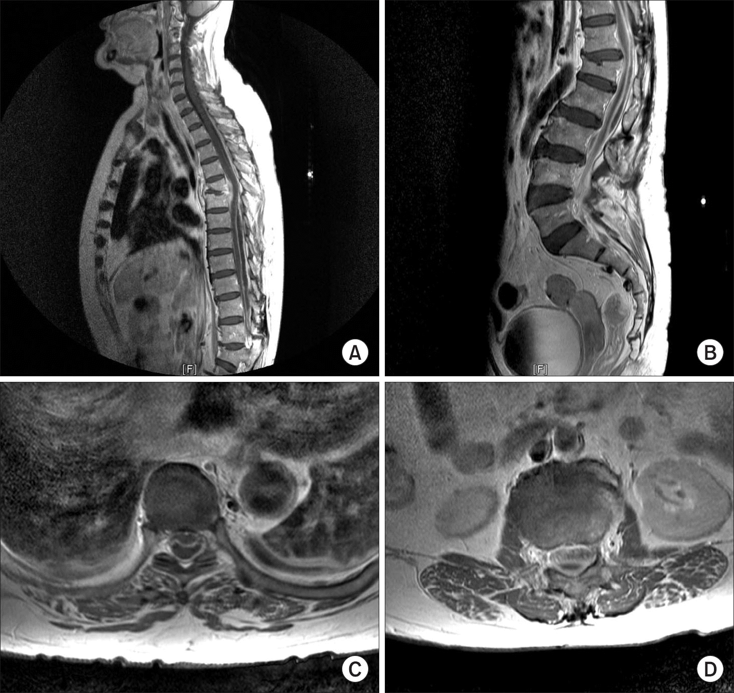
Preoperative sagittal (A) and T8 level axial (C) T1-weighted contrast enhanced cervicothoracic magnetic resonance imaging (MRI) shows T5–6 disc space narrowing, endplate destruction, and epidural swelling with enhancement, suggesting infective spondylodiscitis. It also shows widespread thoracolumbar posterior subdural fluid collection with a rim enhancement, which suggest subdural emphyema. Preoperative sagittal (B) and L2 level axial (D) T1-weighted contrast enhanced lumbar MRI shows widespread thoracolumbar posterior subdural fluid collection with rim enhancement, suggesting subdural emphysema. It also shows clumping of the cauda equina with an enhancement of dural sac, suggesting arachnoiditis.


 PDF
PDF ePub
ePub Citation
 Print
Print


 XML Download