Abstract
Purpose
Materials and Methods
Results
Conclusion
REFERENCES
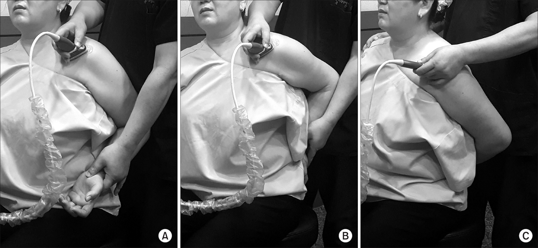
 | Figure 1This figure shows positioning of the arm during sonography. (A) Shoulder extension position. (B) Modified Crass position. (C) Crass position. |
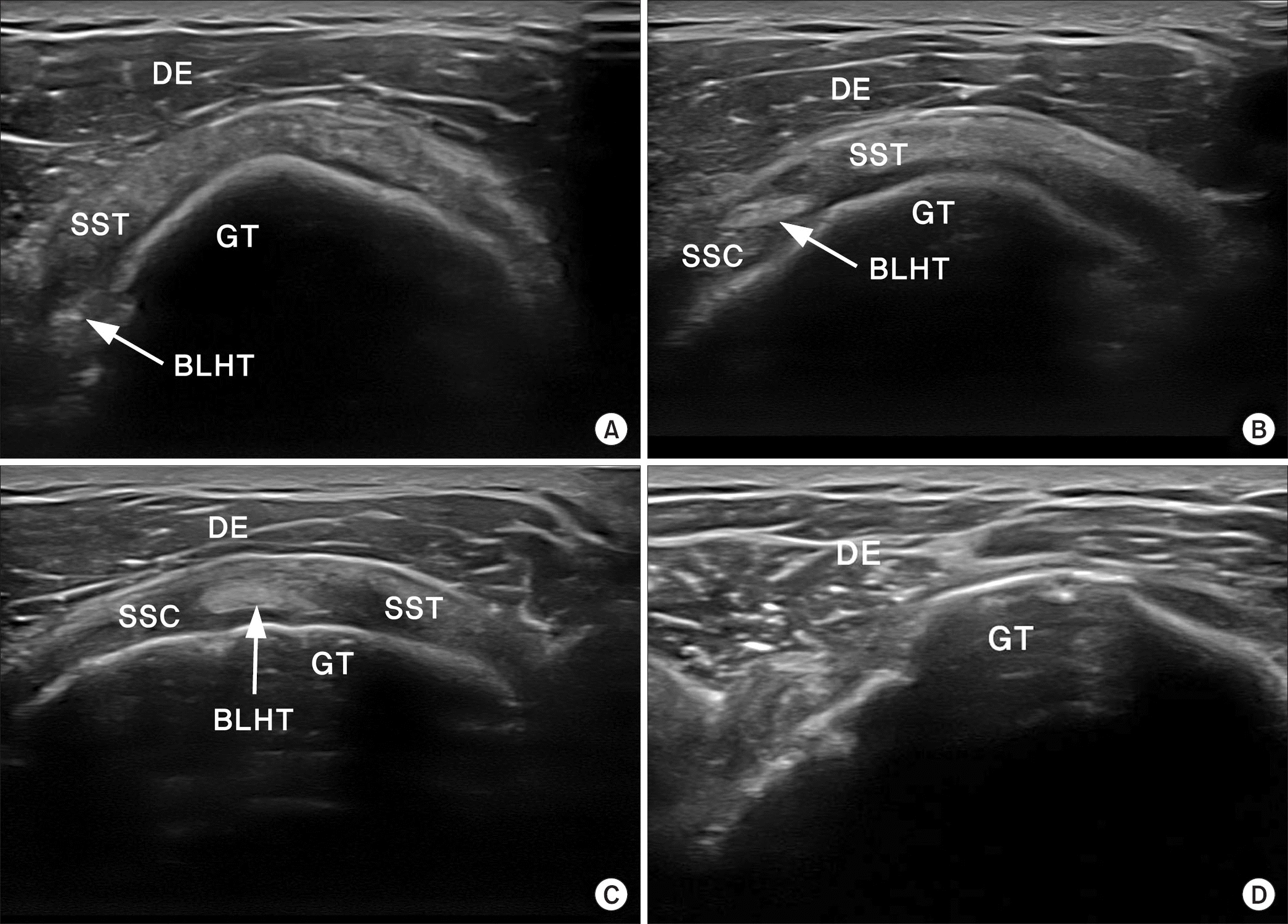
 | Figure 2These sonographic images show the grading of the short-axis view of the rotator interval. (A) Grade I, when the border between the BLHT and the SST tendon is not observed prominently. (B) Grade II, when the border between the BLHT and the SST tendon is observed prominently, while the border between the BLHT and the SSC tendon is not observed prominently. (C) Grade III, when the border between the SSC tendon, the BLHT, and the SST tendon is prominently observed in one field of view. (D) Grade X, when the border is ambiguous due to a complete rupture of the SST tendon or BLHT or an extensive rupture of the rotator cuff. DE, deltoid muscle; SST, supraspinatus; GT, greater tubercle; BLHT, biceps long head tendon; SSC, subscapularis. |
Table 1
Values are presented as number only, mean±standard deviation, or median (range). Group E, shoulder extension position; Group EM, shoulder extension and modified Crass position; Group EMC, shoulder extension, modified Crass position, and Crass position; BMI, body mass index; VAS, visual analogue scale.
Table 2
Table 3
Values are presented as number only. Grade I, when the border between the biceps long head tendon and the supraspinatus tendon is not observed prominently; Grade II, when the border between the biceps long head tendon and the supraspinatus tendon is observed prominently, while the border between the biceps long head tendon and the subscapularis tendon is not observed prominently; Grade III, when tthe border between the subscapularis tendon, the biceps long head tendon, and the supraspinatus tendon is prominently observed in one field of view; Grade X, when the border is ambiguous due to a complete rupture of the supraspinatus tendon or biceps long head tendon or an extensive rupture of the rotator cuff.


 PDF
PDF ePub
ePub Citation
 Print
Print


 XML Download