

 PDF
PDF ePub
ePub Citation
 Print
Print


Abstract
Purpose
The purpose of this study is to evaluate the risk factors for the occurrence of cut-out of proximal femoral nail by a lag screw as the treatment for intertrochanteric fractures.
Materials and Methods
A total of 151 patients (76 males and 75 females; mean age, 73.7±12.1 years), who were diagnosed with intertrochanteric fracture at Gyeongsang National University Hospital between January 2011 and March 2016, with a follow-up of at least for 6 months were included in this retrospective study. Various risk factors, such as demographic data, osteoporosis, collodiaphyseal angle (CDA) (≤130˚ or >130˚), tip-apex distance (TAD) (≤25 mm or >25 mm), and the position of lag screw in the femur head (quadrant) related to the occurrence of cut-out were taken into consideration. The strength of association for each factor was determined through the calculation of the odds ratio (OR), within the 95% confidence interval (CI). First, we performed univariate logistic regression analyses for all variables; then, we performed a multivariate logistic regression analysis, using only the significant variables that had resulted from the univariate analysis.
Results
Among the 151 cases, the occurrence of cut-out was observed in 14 cases (9.3%). In a univariate analysis, the fracture patterns based on the AO/OTA classification (p=0.045), CDA (p<0.001) and the position of lag screw in the femur head (quadrant) (p=0.001) showed a significant association with the occurrence of the cut-out. However, TAD was not significantly associated with the cut-out (p=0.886). Various factors, which were significant in univariate analyses, were included in multivariate analyses. In multivariate analyses, CDA (OR, 12.291; 95% CI, 2.559-59.034; p=0.002), and quadrant (OR, 7.194; 95% CI, 1.712-30.303; p=0.007) were significantly associated with the cut-out.
Go to : 
REFERENCES
1. Hagino H, Yamamoto K, Ohshiro H, Nakamura T, Kishimoto H, Nose T. Changing incidence of hip, distal radius, and proximal humerus fractures in Tottori Prefecture, Japan. Bone. 1999; 24:265–70.

2. Richmond J, Aharonoff GB, Zuckerman JD, Koval KJ. Mortality risk after hip fracture. J Orthop Trauma. 2003; 17:53–6.
3. Koval KJ, Aharonoff GB, Rokito AS, Lyon T, Zuckerman JD. Patients with femoral neck and intertrochanteric fractures. Are they the same? Clin Orthop Relat Res. 1996; 330:166–72.
4. Barton TM, Gleeson R, Topliss C, Greenwood R, Harries WJ, Chesser TJ. A comparison of the long gamma nail with the sliding hip screw for the treatment of AO/OTA 31-A2 fractures of the proximal part of the femur: a prospective randomized trial. J Bone Joint Surg Am. 2010; 92:792–8.
5. Schipper IB, Steyerberg EW, Castelein RM. . Treatment of unstable trochanteric fractures. Randomised comparison of the gamma nail and the proximal femoral nail. J Bone Joint Surg Br. 2004; 86:86–94.
6. Haidukewych GJ, Israel TA, Berry DJ. Reverse obliquity fractures of the intertrochanteric region of the femur. J Bone Joint Surg Am. 2001; 83:643–50.
7. Sadowski C, Lübbeke A, Saudan M, Riand N, Stern R, Hoff-meyer P. Treatment of reverse oblique and transverse intertrochanteric fractures with use of an intramedullary nail or a 95 degrees screw-plate: a prospective, randomized study. J Bone Joint Surg Am. 2002; 84:372–81.
8. Parker MJ, Handoll HH. Gamma and other cephalocondylic intramedullary nails versus extramedullary implants for extracapsular hip fractures in adults. Cochrane Database Syst Rev. 2010; 9:CD000093.
9. Riina J, Tornetta P 3rd, Ritter C, Geller J. Neurologic and vascular structures at risk during anterior-posterior locking of retrograde femoral nails. J Orthop Trauma. 1998; 12:379–81.
10. Mavrogenis AF, Panagopoulos GN, Megaloikonomos PD. . Complications after hip nailing for fractures. Orthopedics. 2016; 39:e108–16.
11. Lorich DG, Geller DS, Nielson JH. Osteoporotic pertrochanteric hip fractures: management and current controversies. Instr Course Lect. 2004; 53:441–54.
12. Kim WY, Han CH, Park JI, Kim JY. Failure of intertrochanteric fracture fixation with a dynamic hip screw in relation to pre-operative fracture stability and osteoporosis. Int Orthop. 2001; 25:360–2.
13. Baumgaertner MR, Curtin SL, Lindskog DM, Keggi JM. The value of the tip-apex distance in predicting failure of fixation of peritrochanteric fractures of the hip. J Bone Joint Surg Am. 1995; 77:1058–64.
14. Wu CC, Shih CH, Chen WJ, Tai CL. Treatment of cutout of a lag screw of a dynamic hip screw in an intertrochanteric fracture. Arch Orthop Trauma Surg. 1998; 117:193–6.
15. Cleveland M, Bosworth DM, Thompson FR, Wilson HJ Jr, Ishizuka T. A ten-year analysis of intertrochanteric fractures of the femur. J Bone Joint Surg Am. 1959; 41:1399–408.
16. Yu J, Zhang C, Li L. . Internal fixation treatments for intertrochanteric fracture: a systematic review and meta-analysis of randomized evidence. Sci Rep. 2015; 5:18195.
17. Shen J, Hu C, Yu S, Huang K, Xie Z. A meta-analysis of percutenous compression plate versus intramedullary nail for treatment of intertrochanteric HIP fractures. Int J Surg. 2016; 29:151–8.
18. Long H, Lin Z, Lu B. . Percutaneous compression plate versus dynamic hip screw for treatment of intertrochanteric hip fractures: a overview of systematic reviews and update meta-analysis of randomized controlled trials. Int J Surg. 2016; 33:1–7.
19. Pajarinen J, Lindahl J, Savolainen V, Michelsson O, Hirven-salo E. Femoral shaft medialisation and neck-shaft angle in unstable pertrochanteric femoral fractures. Int Orthop. 2004; 28:347–53.
20. Hsueh KK, Fang CK, Chen CM, Su YP, Wu HF, Chiu FY. Risk factors in cutout of sliding hip screw in intertrochanteric fractures: an evaluation of 937 patients. Int Orthop. 2010; 34:1273–6.
21. Parker MJ. Valgus reduction of trochanteric fractures. Injury. 1993; 24:313–6.
22. Andruszkow H, Frink M, Frömke C, et al. Tip apex distance, hip screw placement, and neck shaft angle as potential risk factors for cut-out failure of hip screws after surgical treatment of intertrochanteric fractures. Int Orthop. 2012; 36:2347–54.
23. Kuzyk PR, Zdero R, Shah S, Olsen M, Waddell JP, Schemitsch EH. Femoral head lag screw position for cephalomedul-lary nails: a biomechanical analysis. J Orthop Trauma. 2012; 26:414–21.
24. Davis TR, Sher JL, Horsman A, Simpson M, Porter BB, Checketts RG. Intertrochanteric femoral fractures. Mechanical failure after internal fixation. J Bone Joint Surg Br. 1990; 72:26–31.
25. Den Hartog BD, Bartal E, Cooke F. Treatment of the unstable intertrochanteric fracture. Effect of the placement of the screw, its angle of insertion, and osteotomy. J Bone Joint Surg Am. 1991; 73:726–33.
26. Geller JA, Saifi C, Morrison TA, Macaulay W. Tip-apex distance of intramedullary devices as a predictor of cut-out failure in the treatment of peritrochanteric elderly hip fractures. Int Orthop. 2010; 34:719–22.
27. Herman A, Landau Y, Gutman G, Ougortsin V, Chechick A, Shazar N. Radiological evaluation of intertrochanteric fracture fixation by the proximal femoral nail. Injury. 2012; 43:856–63.
28. Mingo-Robinet J, Torres-Torres M, Martínez-Cervell C, et al. Comparative study of the second and third generation of gamma nail for trochanteric fractures: review of 218 cases. J Orthop Trauma. 2015; 29:e85–90.
29. Kraus M, Krischak G, Wiedmann K. . Clinical evaluation of PFNA® and relationship between the tip-apex distance and mechanical failure. Unfallchirurg. 2011; 114:470–8.
30. Kane P, Vopat B, Heard W. . Is tip apex distance as important as we think? A biomechanical study examining optimal lag screw placement. Clin Orthop Relat Res. 2014; 472:2492–8.
Go to :
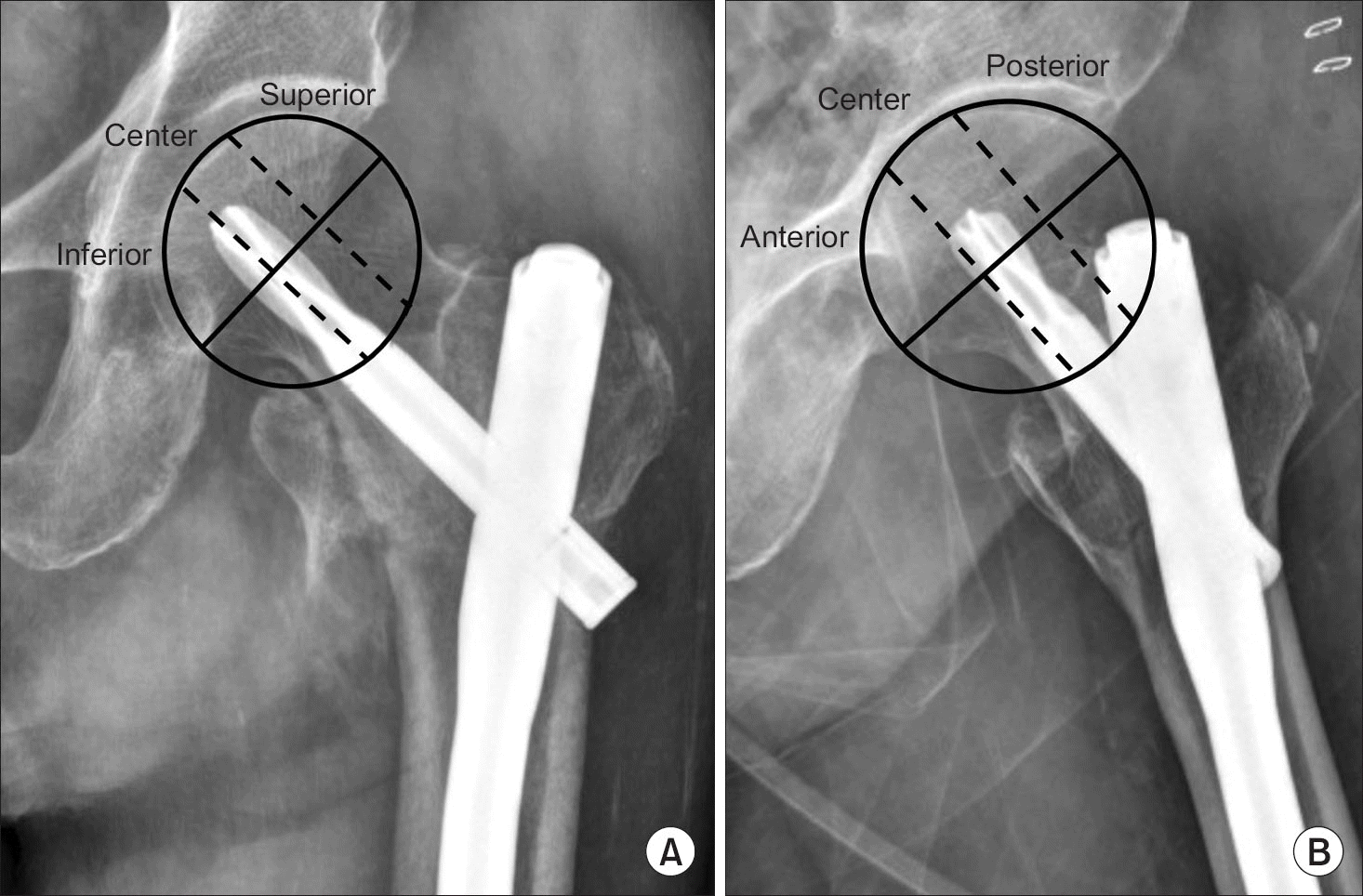
Table 1
Demographic Data of the Patients in Each Group
Table 2
Distribution of Fractures according to AO the Classification*
Table 3
Analysis of Postoperative Neck-Shaft Angle Difference to the Un-Injured Side
| Difference to contralateral side | Total | Cut-out frequency |
|---|---|---|
| <-5° (varus) | 8 | 5 (62.5) |
| -5°–0° | 34 | 4 (11.8) |
| 0°–5° | 53 | 3 (5.7) |
| 5°–10° | 40 | 1 (2.5) |
| >10° (valgus) | 16 | 1 (6.3) |
Table 4
The Statistical Outcomes of the Present Study
 XML Download