

 PDF
PDF ePub
ePub Citation
 Print
Print


Abstract
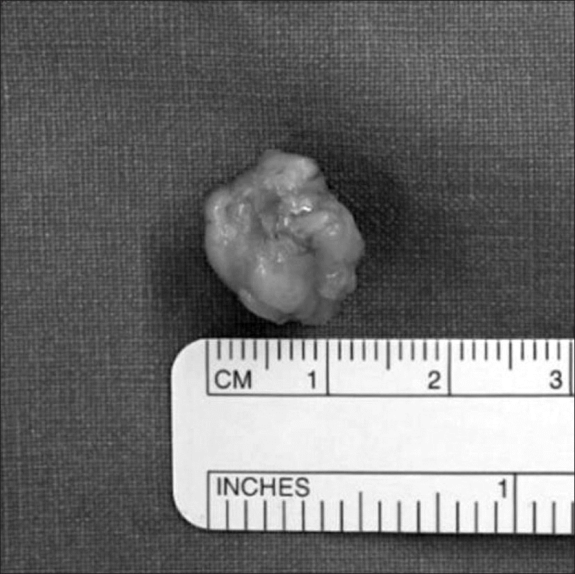
Glomus tumor is a kind of hemangioma that occurs at the glomerulus in the subcutaneous layer. It mainly occurs at the distal hand and subungual area, and rarely at the knee joint. Pain, tenderness, and cold intolerance are known symptoms; however, symptoms in practice are not so easily detectable, and the diagnosis can be delayed if it is presented at areas other than the hand. If the diagnosis is delayed, patients could suffer extreme pain. Therefore, early diagnosis and surgical treatment are important. Ultrasound and magnetic resonance imaging were used to diagnose glomus tumor in our cases, which were found in subcutaneous tissue and muscle fascia. We claim that, for patients with persistent pain, known symptoms—extreme pain, cold intolerance, and tenderness—should be examined carefully and rule out glomus tumor. We report 2 cases of glomus tumors around the knee joint, which is not a common location of occurrence.
Go to : 
REFERENCES
1. Carroll RE, Berman AT. Glomus tumors of the hand: review of the literature and report on twenty-eight cases. J Bone Joint Surg Am. 1972; 54:691–703.
2. Gombos Z, Zhang PJ. Glomus tumor. Arch Pathol Lab Med. 2008; 132:1448–52.

3. Takei TR, Nalebuff EA. Extradigital glomus tumour. J Hand Surg Br. 1995; 20:409–12.
4. Prabhakar S, Dhillon MS, Vasishtha RK, Bali K. Glomus tumor of Hoffa's fat pad and its management by arthroscopic excision. Clin Orthop Surg. 2013; 5:334–7.
5. Kim SH, Suh HS, Choi JH, Sung KJ, Moon KC, Koh JK. Glomus tumor: a clinical and histopathologic analysis of 17 cases. Ann Dermatol. 2000; 12:95–101.
6. Seo BC, Oh DY, Park KS. . Glomus tumor in soleus muscle: a case report. J Korean Soc Plast Reconstr Surg. 2006; 33:518–20.
7. Baek HJ, Lee SJ, Cho KH. . Subungual tumors: clinicopathologic correlation with US and MR imaging findings. Radiographics. 2010; 30:1621–36.
8. Lee MC, Seong SC, Moon YW, Park YK. Glomus tumor adjacent to patellar tendon, a case report. J Korean Knee Soc. 1996; 8:263–7.
9. Lee SH, Lee MC, Seong SC, Jeong GI. Glomus tumor in the infrapatellar fat pad, a case report. J Korean Knee Soc. 2001; 13:236–9.
10. Shaikh R, Alomari AI, Mulliken JB, Fishman SJ, Kozakewich HP, Chaudry G. Subfascial involvement in glomuvenous malformation. Skeletal Radiol. 2014; 43:895–7.
Go to :
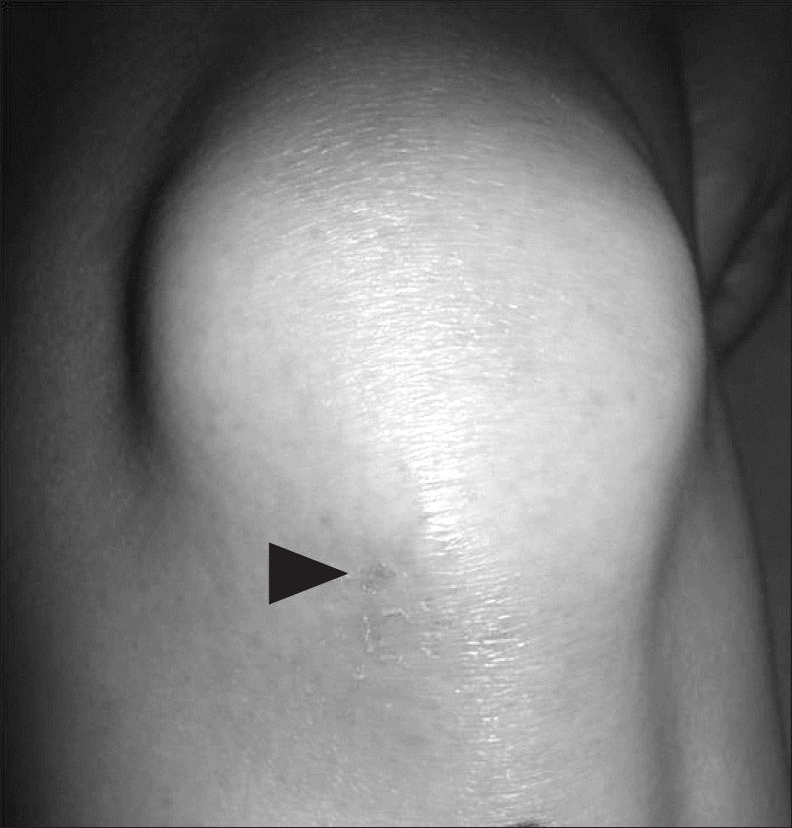 | Figure 1Preoperative clinical photo showing the elevated subcutaneous layer of patellar tendon due to the bluish tender lesion on the left knee (arrowhead). |
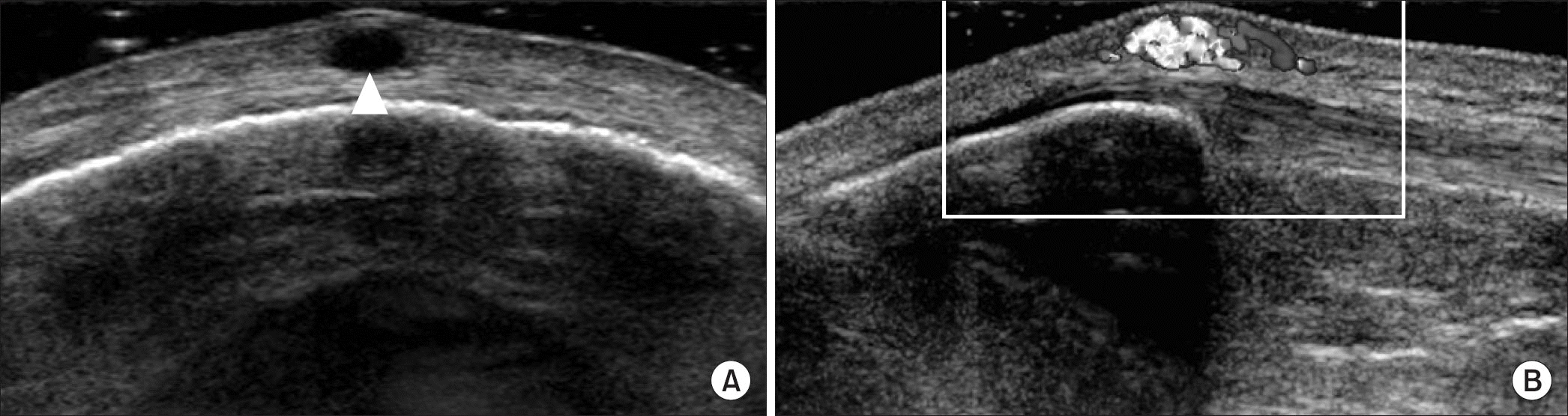 | Figure 2Ultrasound finding of the mass lesion. (A) Well-defined ovoid hypoechoic nodular lesion with a size of about 0.4×0.2×0.5 cm in the subcutaneous fat layer of the prepatellar area on transverse sonograms (arrow). (B) Hypervascular mass lesion on longitudinal Doppler sonograms. |
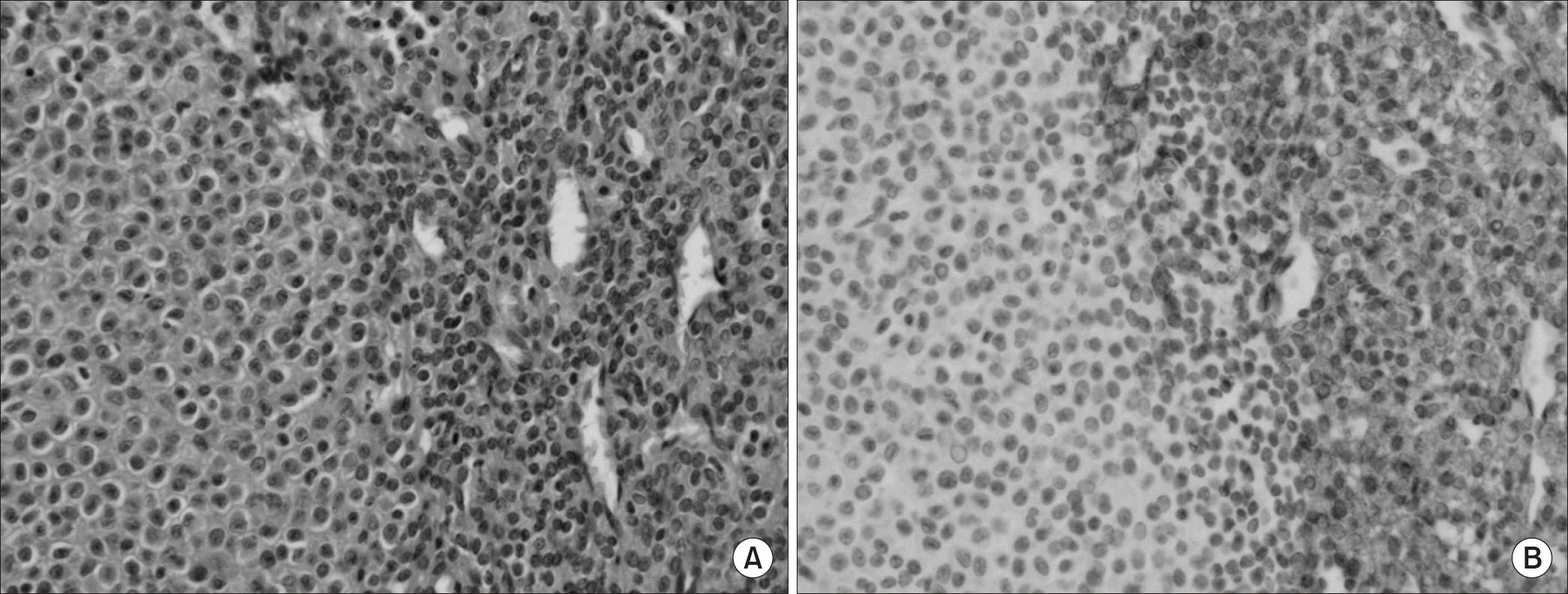
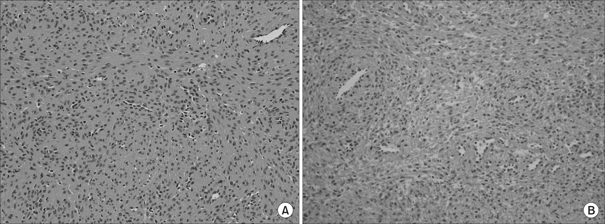 | Figure 3Microscopic findings of the mass lesion. (A) It is composed of capillary sized vessels surrounded by solid proliferation of round-to-cuboidal epithelioid cells with round nuclei (H&E, ×200). (B) Tumor cells are smooth muscle actin (SMA) positive (SMA, ×200). |
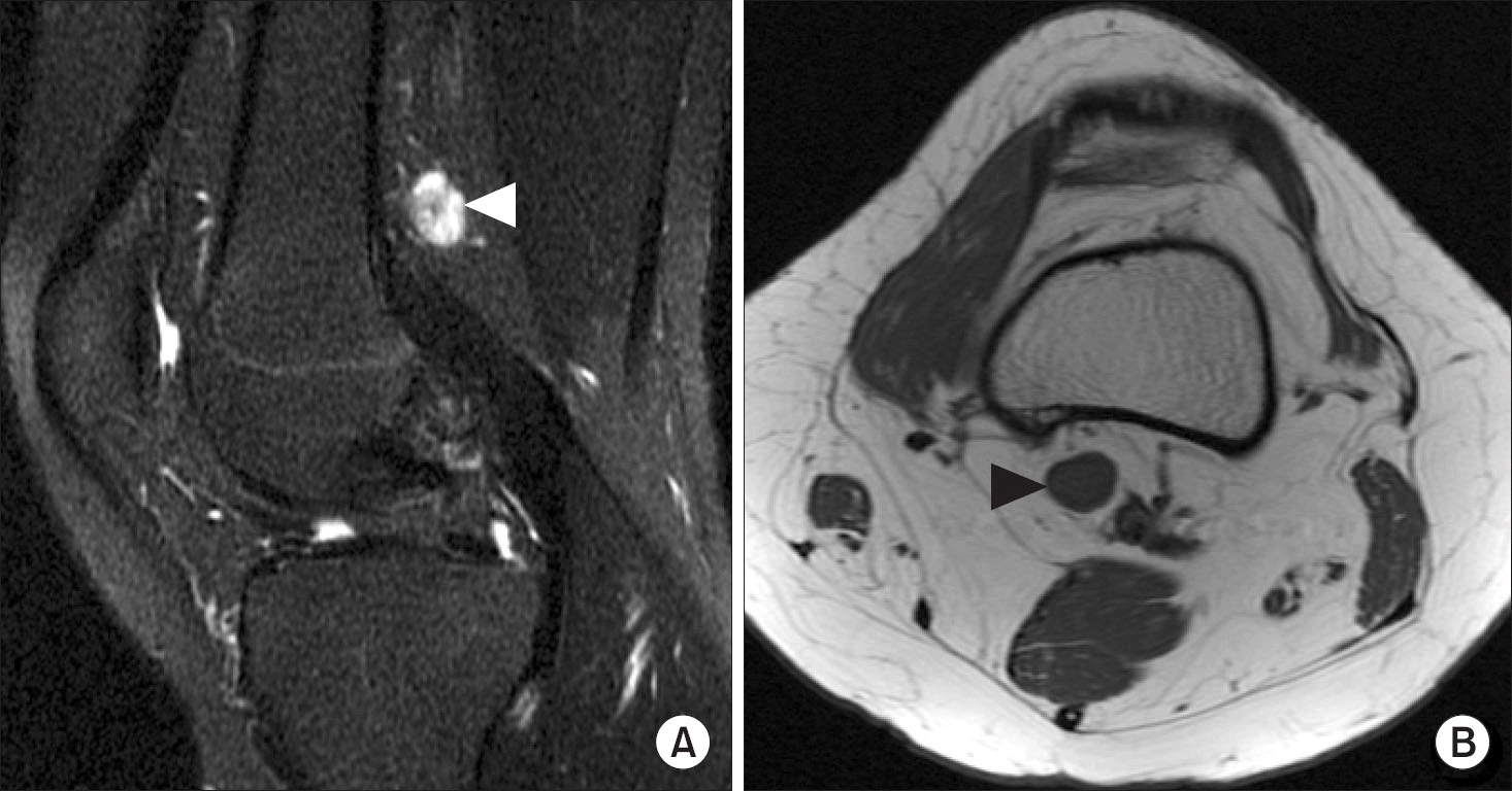 | Figure 4Sagittal T2 (A) and axial T1 (B) magnetic resonance imaging findings. Well-defined 1-cm-sized T1 iso signal (black arrowhead), T2 heterogeneous high signal nodular lesion (white arrowhead) in just the medial and deep aspects of the popliteal artery at the level of femoral insertion site of medial head of gastrocnemius tendon. Separated from neurovascular bundle in popliteal fossa. |
 XML Download