

 PDF
PDF ePub
ePub Citation
 Print
Print


Abstract
Purpose
This study aimed to investigate the clinical features of congenital postural deformities and lower extremity asymmetry with respect to the presence of developmental dysplasia of the hip (DDH) in infants with a discrepancy of the limb length referred for suspected DDH.
Materials and Methods
We retrospectively reviewed the medical records and radiographs of 150 infants who visited Korea University Anam Hospital Orthopedic Clinic for suspected DDH between March 2013 and March 2015.
Results
There were greater numbers of infants with a shorter lower extremity on the left side (n=86, 57.3%) than the right. Plagiocephaly was present in 62 infants and trunk curvature in 124 infants (82.7%). Pelvic tilting—indirectly assessed by a skewed direction of the external genitalia in female infants—was present in 62 infants (63.3%). None of the 139 infants with normal physical examination of the hip were diagnosed with DDH. Of those 11 infants with abnormal findings from the hip physical examination, a total of 6 infants were diagnosed with DDH.
Conclusion
Regardless of the associated findings of congenital postural deformation, all infants diagnosed with DDH had abnormal findings from the physical examination of the hip joint. Thus, we conclude that the hip examination is important as the primary clinical screening in aiding the diagnosis of DDH.
Figures and Tables
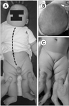 | Figure 1(A) Right sided truncal curvature and shortened left leg is apparent in a 4-month-old boy. (B) Plagiocephaly is apparent with asymmetric flattening of the occiput either on the right or left. (C) The genital direction may indicate pelvic obliquity in girl.
|
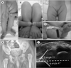 | Figure 2(A) A 4-month-old girl diagnosed as developmental dysplasia of the hip (DDH) with left leg shortening. (B) Galeazzi sign showing limb-length discrepancy was present. (C) Elevated left pelvic obliquity was suspected due to genital deviation to the left. (D) Limited range of hip abduction with inguinal fold asymmetry was seen. (E) Plain radiograph revealed DDH on the left side. (F) On ultrasonography, left hip dysplasia Graf type IV was shown with an α angle of 41° and a β angle of 55°.
|

References
1. Choi IH, Chung CY, Cho TJ, Yoo WJ, Park MS, Lee DY. Pediatric orthopaedics. 3rd ed. Seoul: Koonja;2009. p. 340–350.
2. Fitch RD. Ultrasound for screening and management of developmental dysplasia of the hip. N C Med J. 2014; 75:142–145.

3. Good C, Walker G. Moulded baby syndrome and unilateral “tight” hips. Br Med J (Clin Res Ed). 1983; 287:1675–1676.
4. Good C, Walker G. The hip in the moulded baby syndrome. J Bone Joint Surg Br. 1984; 66:491–492.
5. Klaue K, Durnin CW, Ganz R. The acetabular rim syndrome. A clinical presentation of dysplasia of the hip. J Bone Joint Surg Br. 1991; 73:423–429.
6. Sewell MD, Eastwood DM. Screening and treatment in developmental dysplasia of the hip-where do we go from here? Int Orthop. 2011; 35:1359–1367.
7. Shorter D, Hong T, Osborn DA. Screening programmes for developmental dysplasia of the hip in newborn infants. Cochrane Database Syst Rev. 2011; (9):CD004595.
8. Talbot CL, Paton RW. Screening of selected risk factors in developmental dysplasia of the hip: an observational study. Arch Dis Child. 2013; 98:692–696.
9. Cha SM, Shin HD, Lee SH, Kim BK. An analysis of cases referred from the primary healthcare institution with suspected developmental dysplasia of the hip: a prospective study. J Korean Orthop Assoc. 2011; 46:501–506.
10. Narayanan U, Mulpuri K, Sankar WN, Clarke NM, Hosalkar H, Price CT. Reliability of a new radiographic classification for developmental dysplasia of the hip. J Pediatr Orthop. 2015; 35:478–484.
11. Rubio AS, Griffet JR, Caci H, Bérard E, El Hayek T, Boutté P. The moulded baby syndrome: incidence and risk factors regarding 1,001 neonates. Eur J Pediatr. 2009; 168:605–611.
12. Watson GH. Relation between side of plagiocephaly, dislocation of hip, scoliosis, bat ears, and sternomastoid tumours. Arch Dis Child. 1971; 46:203–210.
13. Hamanishi C, Tanaka S. Turned head--adducted hip--truncal curvature syndrome. Arch Dis Child. 1994; 70:515–519.
14. Seringe R, Langlais J, Bonnet JC. Congenital asymmetrical pelvis. Clinical, radiological study and outcome. Rev Chir Orthop Reparatrice Appar Mot. 1992; 78:65–73.
15. Screening for the detection of congenital dislocation of the hip. Arch Dis Child. 1986; 61:921–926.
16. Paton RW, Choudry Q. Neonatal foot deformities and their relationship to developmental dysplasia of the hip: an 11-year prospective, longitudinal observational study. J Bone Joint Surg Br. 2009; 91:655–658.
17. Graf R. The diagnosis of congenital hip-joint dislocation by the ultrasonic Combound treatment. Arch Orthop Trauma Surg. 1980; 97:117–133.
18. Graf R. Hip sonography: diagnosis and managment of infant hip dysplasia. Berlin: Springer-Verlag Berlin Heidelberg;2006.
19. Rosendahl K, Markestad T, Lie RT. Ultrasound in the early diagnosis of congenital dislocation of the hip: the significance of hip stability versus acetabular morphology. Pediatr Radiol. 1992; 22:430–433.
20. Holen KJ, Tegnander A, Bredland T, et al. Universal or selective screening of the neonatal hip using ultrasound? A prospective, randomised trial of 15,529 newborn infants. J Bone Joint Surg Br. 2002; 84:886–890.
21. Rosendahl K, Markestad T, Lie RT. Ultrasound screening for developmental dysplasia of the hip in the neonate: the effect on treatment rate and prevalence of late cases. Pediatrics. 1994; 94:47–52.
22. Omeroğlu H, Koparal S. The role of clinical examination and risk factors in the diagnosis of developmental dysplasia of the hip: a prospective study in 188 referred young infants. Arch Orthop Trauma Surg. 2001; 121:7–11.
23. Buxton RA, Macnicol MF. Infantile skeletal skew: the use of ultrasound in management. J Pediatr Orthop B. 2004; 13:75–80.
24. Dunn PM. Congenital postural deformities. Br Med Bull. 1976; 32:71–76.
25. Lloyd-roberts GC, Pilcher MF. Structural idiopathic scoliosis in infancy: a study of the natural history of 100 patients. J Bone Joint Surg Br. 1965; 47:520–523.
26. Green NE, Griffin PP. Hip dysplasia associated with abduction contracture of the contralateral hip. J Bone Joint Surg Am. 1982; 64:1273–1281.
27. Ando M, Gotoh E. Significance of inguinal folds for diagnosis of congenital dislocation of the hip in infants aged three to four months. J Pediatr Orthop. 1990; 10:331–334.
28. Jones DA. Importance of the clicking hip in screening for congenital dislocation of the hip. Lancet. 1989; 1:599–601.
29. Paton RW. Screening for hip abnormality in the neonate. Early Hum Dev. 2005; 81:803–806.
30. Jari S, Paton RW, Srinivasan MS. Unilateral limitation of abduction of the hip. A valuable clinical sign for DDH? J Bone Joint Surg Br. 2002; 84:104–107.
 XML Download