

 PDF
PDF ePub
ePub Citation
 Print
Print


Abstract
Purpose
The purpose of this study was to compare magnetic resonance imaging (MRI) and ultrasonography measurement of peri-lumbar
muscle atrophy which is thought to be a cause of low back pain.
Materials and Methods
Eighty-two patients (44 males, 38 females) who visited Wonkang University Hospital from March, 2015 to August, 2015 complaining of lumbar back pain and underwent lumbar MRI were enrolled in this study. Cross section area (CSAMRI) and muscle thickness (MTMRI) of psoas major (PS) and lumbar extensor (LM) located on both sides of L4/5 and L3/4 was measured by MRI, and sono measurement of thickness of the same muscle (MTUS) at the same level of that MRI measurement were analyzed.
Results
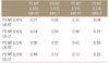
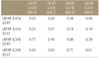
In correlation analysis of PS CSAMRI and PS MTUS is the correlation coefficient of L4/5 was 0.136 (p=0.64), L3/4 right (Rt) was 0.070 (p=0.81), and L3/4 left (Lt) was 0.288 (p=0.32). PS CSAMRI at L4/5 Rt showed that correlation coefficient of PS MTUS showed a positive correlation to 0.559 (p=0.04). In analysis of the PS MTMRI and PS MTUS, the correlation coefficient of L4/5 Rt was measured by a 0.316 (p=0.27), L4/5 Lt was 0.022 (p=0.94), L3/4 Rt was 0.236 (p=0.41), and L3/4 Lt did not show a significant result with 0.287 (p=0.31). In the results of correlation analysis of the LM MTMRI and LM MTUS, the correlation coefficient of L4/5 Rt was 0.207 (p=0.49), L4/5 Lt was 0.051 (p=0.86), and L3/4 was Rt 0.048 (p=0.87), L3/4 Lt did not show a significant value with 0.154 (p=0.61).
Figures and Tables
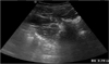
 | Figure 1Measurement of the multifidus muscle thickness between subcutaneous layer and muscle layer on the longitudinal scan.
|
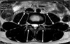
 | Figure 2Measurement of the psoas muscle thickness between the surface and deep connections on the transverse scan.
|
 | Figure 3This shows the thickness of psoas major muscle and multifidus muscle and the cross sectional areas of psoas major muscle.
|


References
1. Kirchmair L, Entner T, Wissel J, Moriggl B, Kapral S, Mitterschiffthaler G. A study of the paravertebral anatomy for ultrasound-guided posterior lumbar plexus block. Anesth Analg. 2001; 93:477–481.

2. Nofsinger C, Konin JG. Diagnostic ultrasound in sports medicine: current concepts and advances. Sports Med Arthrosc. 2009; 17:25–30.
3. Hodges PW, Pengel LH, Herbert RD, Gandevia SC. Measurement of muscle contraction with ultrasound imaging. Muscle Nerve. 2003; 27:682–692.
4. McMeeken JM, Beith ID, Newham DJ, Milligan P, Critchley DJ. The relationship between EMG and change in thickness of transversus abdominis. Clin Biomech (Bristol, Avon). 2004; 19:337–342.
5. Bunce SM, Moore AP, Hough AD. M-mode ultrasound: a reliable measure of transversus abdominis thickness? Clin Biomech (Bristol, Avon). 2002; 17:315–317.
6. Kiesel KB, Uhl TL, Underwood FB, Rodd DW, Nitz AJ. Measurement of lumbar multifidus muscle contraction with rehabilitative ultrasound imaging. Man Ther. 2007; 12:161–166.
7. Van K, Hides JA, Richardson CA. The use of real-time ultrasound imaging for biofeedback of lumbar multifidus muscle contraction in healthy subjects. J Orthop Sports Phys Ther. 2006; 36:920–925.
8. Kamaz M, Kireşi D, Oğuz H, Emlik D, Levendoğlu F. CT measurement of trunk muscle areas in patients with chronic low back pain. Diagn Interv Radiol. 2007; 13:144–148.
9. Shim DM, Kim TK, Lee SJ, Song SY. Comparison of ultrasonography and MRI in measuring of cervical soft tissue structure. J Korean Orthop Assoc. 2011; 46:282–287.
10. Richardson C, Jull G, Hodges P, Hides J, Panjabi MM. Therapeutic exercise for spinal segmental stabilization in low back pain: scientific basis and clinical approach. Edinburgh: Churchill Livingstone;1999.
11. Kay AG. An extensive literature review of the lumbar multifidus: anatomy. J Man Manip Ther. 2000; 8:102–114.
12. Arbanas J, Klasan GS, Nikolic M, Jerkovic R, Miljanovic I, Malnar D. Fibre type composition of the human psoas major muscle with regard to the level of its origin. J Anat. 2009; 215:636–641.
13. Stokes M, Rankin G, Newham DJ. Ultrasound imaging of lumbar multifidus muscle: normal reference ranges for measurements and practical guidance on the technique. Man Ther. 2005; 10:116–126.
14. Macintosh JE, Valencia F, Bogduk N, Munro RR. The morphology of the human lumbar multifidus. Clin Biomech (Bristol, Avon). 1986; 1:196–204.
15. Bogduk N, Macintosh JE, Pearcy MJ. A universal model of the lumbar back muscles in the upright position. Spine (Phila Pa 1976). 1992; 17:897–913.
16. Hides JA, Cooper DH, Stokes MJ. Diagnostic ultrasound imaging for measurement of the lumbar multifidus muscle in normal young adults. Physiother Theory Pract. 1992; 8:19–26.
17. Hoshikawa Y, Muramatsu M, Iida T, et al. Influence of the psoas major and thigh muscularity on 100-m times in junior sprinters. Med Sci Sports Exerc. 2006; 38:2138–2143.
18. Hashimoto BE, Kramer DJ, Wiitala L. Applications of musculoskeletal sonography. J Clin Ultrasound. 1999; 27:293–318.
19. Stokes M, Hides JA, Nassiri DK. Musculoskeletal ultrasound imaging: Diagnostic and treatment aid in rehabilitation. Phys Ther Rev. 1997; 2:73–92.
20. Iannotti JP, Ciccone J, Buss DD, et al. Accuracy of officebased ultrasonography of the shoulder for the diagnosis of rotator cuff tears. J Bone Joint Surg Am. 2005; 87:1305–1311.
21. Hides JA, Richardson CA, Jull GA. Multifidus muscle recovery is not automatic after resolution of acute, first-episode low back pain. Spine (Phila Pa 1976). 1996; 21:2763–2769.
22. Shim DM, Kim TK, Oh SK, Lee SJ, Yang HS. Comparison of ultrasonography and magnetic resonance imaging in measurement of lumbar spine anatomic structures. J Korean Orthop Assoc. 2012; 47:140–145.
23. Takai Y, Katsumata Y, Kawakami Y, Kanehisa H, Fukunaga T. Ultrasound method for estimating the cross-sectional area of the psoas major muscle. Med Sci Sports Exerc. 2011; 43:2000–2004.
24. Lee JP, Tseng WY, Shau YW, Wang CL, Wang HK, Wang SF. Measurement of segmental cervical multifidus contraction by ultrasonography in asymptomatic adults. Man Ther. 2007; 12:286–294.
 XML Download