

 PDF
PDF ePub
ePub Citation
 Print
Print


Abstract
Schwannoma is the most common benign tumor of peripheral nerves and usually appears on the trunk, head and neck, or extremities. A mass arising at popliteal fossa can be misdiagnosed as a popliteal cyst. We report on a rare case of a popliteal schwannoma mimicking a popliteal cyst in a 39-year-old female who showed a clinical presentation similar to that of a popliteal cyst. Diagnosis was delayed until ultrasonographic evaluation was performed due to its anatomical location, the same as that of a popliteal cyst. We describe the clinical significance and ultrasonographic findings of the schwannoma for initial differential diagnosis from a popliteal cyst.
Figures and Tables
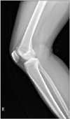
Figure 2
Ultrasonographic findings showing a well-defined, hypoechoic oval shaped nodular mass filled with nonhomogeneous components (A) and tibial nerve fascicular continuity at its proximal end (B). (C) Compression of the popliteal vein was observed at the bottom of the mass.

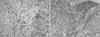
Figure 3
Magnetic resonance imaging findings of the mass. Saggital T1- (A), T2- (B), and axial T2 contrast enhanced-weighted images demonstrating a well-marginated mass with hypointense (A), hyperintense (B), inner heterogeneous (C) signals, respectively.
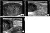
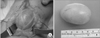
Figure 4
Photograph of the right popliteal fossa mass at surgery, which confirmed a well-defined mass originating from the tibial nerve branch (A) and an enucleated mass, 5×4×3 cm in size (B).

References
1. Kim JI, Kim UJ, Moon TY, Lee IS, Song YS, Choi KU. Diagnostic value of MRI in schwannoma. J Korean Bone Joint Tumor Soc. 2014; 20:60–65.

2. Komurcu E, Golge UH, Kaymaz B, Erdogan N. Popliteal schwannoma mimicking baker cyst: an unusual case. J Surg Case Rep [Internet]. 2013. cited 2013 Aug 29. DOI: 10.1093/jscr/rjt066. Available from: http://jscr.oxfordjournals.org/content/2013/8/rjt066.long.
3. Shariq O, Radha S, Konan S. Common peroneal nerve schwannoma: an unusual differential for a symptomatic knee lump. BMJ Case Rep [Internet]. 2012. cited 2012 Dec 3. DOI: 10.1136/bcr-2012-007346. Available from: http://casereports.bmj.com/content/2012/bcr-2012-007346.long.
4. Andrychowski J, Czernicki Z, Jasielski P. Schwannoma of the common peroneal nerve. A differential diagnosis versus rare popliteal cyst. Neurol Neurochir Pol. 2012; 46:396–400.
5. Maraziotis T, Panagiotopoulos E, Panagiotopoulos V, Panagiotopoulos K. Neurilemoma of the popliteal fossa: report of two cases with long subclinical course and misleading presentation. Acta Orthop Belg. 2005; 71:496–499.
6. Green DP, Hotchkiss RN, Pederson WC, Wolfe SW. Green's operative hand surgery. 5th ed. Philadelphia: Churchill Livingstone;2005. p. 2211–2264.
7. King AD, Ahuja AT, King W, Metreweli C. Sonography of peripheral nerve tumors of the neck. AJR Am J Roentgenol. 1997; 169:1695–1698.
8. Simonovský V. Peripheral nerve schwannoma preoperatively diagnosed by sonography: report of three cases and discussion. Eur J Radiol. 1997; 25:47–51.
9. Stull MA, Moser RP Jr, Kransdorf MJ, Bogumill GP, Nelson MC. Magnetic resonance appearance of peripheral nerve sheath tumors. Skeletal Radiol. 1991; 20:9–14.
10. Suh JS, Abenoza P, Galloway HR, Everson LI, Griffiths HJ. Peripheral (extracranial) nerve tumors: correlation of MR imaging and histologic findings. Radiology. 1992; 183:341–346.
 XML Download