

 PDF
PDF ePub
ePub Citation
 Print
Print


Abstract
Purpose
Spinopelvic dissociation which occurs by high energy trauma with associated fractures is rare. Treatment is difficult and only a few studies on treatment of spinopelvic dissociation have been reported. Therefore we evaluated spinopelvic dissociation patients treated with iliac screw.
Materials and Methods
We analyzed patients who underwent surgery using an iliac screw from 2005 to 2010. Preoperative radiologic classification was performed using the level of the transverse fracture line of the sacrum, shape of the fracture, and Roy-Camille classification. Neurologic evaluation was performed using Gibbons classification. Eleven patients underwent surgery with a pedicle screw in 1 level (L5 to S1) and bilateral iliac screws were added.
Results
A total of 11 patients were included in this study. The level of the transverse fracture line of the sacrum was mainly at S2, and there were mostly type 3 or 4 in Roy-Camille classification. Bony union was checked in 11 patients without metal failure. Six of 7 patients were treated by posterior decompression. Among them, 5 patients recovered from neurological deficit and 1 patient still had a sensory disorder on both lower legs.
Conclusion
The more displacement of fracture, the more neurologic deficit occurred. In addition, we think that aggressive surgical treatment for spinopelvic dissociation should be considered, because a good clinical result was achieved with 1 level (L5 to S1) fixation and bilateral iliac screw fixation.
Figures and Tables
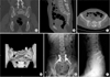 | Figure 1A 44-year-old-female suffered spinopelvic dissociation. Preoperative computed tomography, coronal image (A), sagittal image (B), axial image (C), three-dimensional computed tomography reconstruction image (D). (E) Postoperative anteroposterior radiograph after lumbopelvic fixation with iliac screw. (F) Postoperative lateral radiograph after lumbopelvic fixation with iliac screw.
|
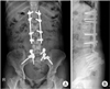 | Figure 2A 30-year-old-female suffered spinopelvic dissociation. (A) At 7-year postoperative, anteroposterior radiographs. (B) At 7-year postoperative, lateral radiographs.
|
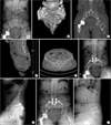 | Figure 3A 66-year-old-female suffered spinopelvic dissociation and was treated 47 days after the injury. Preoperative radiograph (A), three dimensional computed tomography reconstruction image (B), coronal reconstruction image (C), sagittal reconstruction image (D), and axial reconstruction image (E). Postoperative anteroposterior radiograph (F) and lateral radiograph (G) after lumbopelvic fixation. At 1-year postoperative, anteroposterior radiograph (H), and lateral radiograph (I).
|
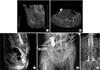 | Figure 4A 42-year-old-male suffered spinopelvic dissociation with pelvic ring injury and T12 bursting fracture. Preoperative computed tomography, sagittal image (A), and axial image (B). (C) Preoperative magnetic resonance imaging sagittal image. (D) External fixator application for pelvic ring fracture. (E) Postoperative anteroposterior radiograph after lumbopelvic fixation with iliac screw.
|

References
1. Ebraheim NA, Biyani A, Salpietro B. Zone III fractures of the sacrum. A case report. Spine (Phila Pa 1976). 1996; 21:2390–2396.
2. Gribnau AJ, van Hensbroek PB, Haverlag R, Ponsen KJ, Been HD, Goslings JC. U-shaped sacral fractures: surgical treatment and quality of life. Injury. 2009; 40:1040–1048.

3. Schildhauer TA, Ledoux WR, Chapman JR, Henley MB, Tencer AF, Routt ML Jr. Triangular osteosynthesis and iliosacral screw fixation for unstable sacral fractures: a cadaveric and biomechanical evaluation under cyclic loads. J Orthop Trauma. 2003; 17:22–31.
4. Nork SE, Jones CB, Harding SP, Mirza SK, Routt ML Jr. Percutaneous stabilization of U-shaped sacral fractures using iliosacral screws: technique and early results. J Orthop Trauma. 2001; 15:238–246.
5. Schildhauer TA, Bellabarba C, Nork SE, Barei DP, Routt ML Jr, Chapman JR. Decompression and lumbopelvic fixation for sacral fracture-dislocations with spino-pelvic dissociation. J Orthop Trauma. 2006; 20:447–457.
6. Denis F, Davis S, Comfort T. Sacral fractures: an important problem. Retrospective analysis of 236 cases. Clin Orthop Relat Res. 1988; 227:67–81.
7. Fountain SS, Hamilton RD, Jameson RM. Transverse fractures of the sacrum. A report of six cases. J Bone Joint Surg Am. 1977; 59:486–489.
8. Kim MY, Reidy DP, Nolan PC, Finkelstein JA. Transverse sacral fractures: case series and literature review. Can J Surg. 2001; 44:359–363.
9. Roy-Camille R, Saillant G, Gagna G, Mazel C. Transverse fracture of the upper sacrum. Suicidal jumper's fracture. Spine (Phila Pa 1976). 1985; 10:838–845.
10. Strange-Vognsen HH, Lebech A. An unusual type of fracture in the upper sacrum. J Orthop Trauma. 1991; 5:200–203.
11. Fisher RG. Sacral fracture with compression of cauda equina: surgical treatment. J Trauma. 1988; 28:1678–1680.
12. Gibbons KJ, Soloniuk DS, Razack N. Neurological injury and patterns of sacral fractures. J Neurosurg. 1990; 72:889–893.
13. Hessmann MH, Rommens PM. Transverse fracture-dislocation of the sacrum: a diagnostic pitfall and a surgical challenge. Acta Chir Belg. 2002; 102:46–51.
14. Schildhauer TA, Josten C, Muhr G. Triangular osteosynthesis of vertically unstable sacrum fractures: a new concept allowing early weight-bearing. J Orthop Trauma. 1998; 12:307–314.
15. Hunt N, Jennings A, Smith M. Current management of U-shaped sacral fractures or spino-pelvic dissociation. Injury. 2002; 33:123–126.
16. Robles LA. Transverse sacral fractures. Spine J. 2009; 9:60–69.
17. Taguchi T, Kawai S, Kaneko K, Yugue D. Operative management of displaced fractures of the sacrum. J Orthop Sci. 1999; 4:347–352.
18. Ebraheim NA, Coombs R, Hoeflinger MJ, Zeman C, Jackson WT. Anatomical and radiological considerations in compressive bar technique for posterior pelvic disruptions. J Orthop Trauma. 1991; 5:434–438.
19. Vilela MD, Jermani C, Braga BP. Lumbopelvic fixation and sacral decompression for a U-shaped sacral fracture: case report. Arq Neuropsiquiatr. 2007; 65:865–868.
20. Bellabarba C, Schildhauer TA, Vaccaro AR, Chapman JR. Complications associated with surgical stabilization of high-grade sacral fracture dislocations with spino-pelvic instability. Spine (Phila Pa 1976). 2006; 31:S80–S88.
21. Mouhsine E, Wettstein M, Schizas C, et al. Modified triangular posterior osteosynthesis of unstable sacrum fracture. Eur Spine J. 2006; 15:857–863.
22. Schildhauer TA, McCulloch P, Chapman JR, Mann FA. Anatomic and radiographic considerations for placement of transiliac screws in lumbopelvic fixations. J Spinal Disord Tech. 2002; 15:199–205.
23. Yi C, Hak DJ. Traumatic spinopelvic dissociation or U-shaped sacral fracture: a review of the literature. Injury. 2012; 43:402–408.
24. Tian X, Li J, Sheng W, et al. Morphometry of iliac anchorage for transiliac screws: a cadaver and CT study of the Eastern population. Surg Radiol Anat. 2010; 32:455–462.
25. Jones CB, Sietsema DL, Hoffmann MF. Can lumbopelvic fixation salvage unstable complex sacral fractures? Clin Orthop Relat Res. 2012; 470:2132–2141.
26. Yu BS, Zhuang XM, Zheng ZM, Li ZM, Wang TP, Lu WW. Biomechanical advantages of dual over single iliac screws in lumbo-iliac fixation construct. Eur Spine J. 2010; 19:1121–1128.
27. Soultanis K, Karaliotas GI, Mastrokalos D, Sakellariou VI, Starantzis KA, Soucacos PN. Lumbopelvic fracture-dislocation combined with unstable pelvic ring injury: one stage stabilisation with spinal instrumentation. Injury. 2011; 42:1179–1183.
28. Avadhani A, Shetty AP, Rajasekaran S. Pediatric transverse sacral fracture with cauda equina syndrome. Spine J. 2010; 10:e10–e13.
 XML Download