

 PDF
PDF ePub
ePub Citation
 Print
Print


Since the description of meniscal ossicle was introduced by Wsaotn-Jones and Roberts1) in 1934, a number of authors have reported this rare abnormality. The prevalence of meniscal ossicle was found to be 0.15% among 1,287 consecutive knee magnetic resonance imaging (MRI) examinations.2) The most common site of meniscal ossic le is in the posterior horn of the medial meniscus.3) Traditionally, arthroscopic resection of the ossicle is recommended for symptomatic cases.4) However, some articles recommend arthroscopic resection and meniscal repair as adequate treatments.56) We performed arthroscopic meniscal resection including the ossicle and microafrcture to treat meniscal ossicle and the cartilage lesion in the medial femoral condyle accompanied by medial meniscus posterior horn radial tear. This patient was diagnosed with meniscal ossicle of the knee based on the results of a histopathological biopsy
CASE REPORT
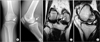
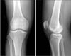
A 48-year-old male was referred to our hospital for pain and intermittent locking symptoms in the right knee that had begun 6 months before. He was treated conservatively at a local clinic, but his symptoms did not improve. Physical examination at our hospital revealed direct tenderness along the right posteromedial joint line and limitation of full flexion because of the pain. The McMurray test was positive. A plain radiograph of the knee revealed a solitary and well-defined oval-shaped bony structure in the posteromedial compartment of the knee (Fig. 1A, 1B). Subsequent MRI revealed that an ossicle with an isointensity cortical rim was embedded within the posterior horn of the medial meniscus and that the posterior horn was detached from its tibial insertion (Fig. 1C, 1D). Arthroscopy was performed with 90-degree flexion of the right left knee. Probing in the arthroscopic view revealed a tear in the posterior horn of the medial meniscus at the tibial insertion. Integrity was partially maintained, but it was loose and unstable because of the degenerative changes in the soft tissue (Fig. 2A). Meanwhile, although the normal meniscus lateral to the tear displayed swelling and a palpable solid mass inside, there were no lesions that suggested an avulsion fracture of the tibial insertion (Fig. 2B). In the meantime, the outerbridge classification grade IV cartilage defect on the medial femoral condyle was observed, likely caused by repeated contact with the enlarged hard posterior horn (Fig. 2C). Meniscectomy was performed accordingly for the posterior horn radial tear, and the mass embedded inside the meniscus was resected along with normal tissue because separate resection was impossible. Suture was not performed because probing after resection showed that reattachment of the residual meniscus to its normal anatomical location was impossible. Meanwhile, marrow stimulation by microfracture was performed for the cartilage lesion in the medial femoral condyle. The resected mass was sized 10×5×10 mm macroscopically and was surrounded by smooth, white cartilage (Fig. 3A). A histological examination revealed mature cancellous bone surrounded by a fibrocartilage layer. There were no signs of mucoid degeneration, revascularization, fibroblastic proliferation or metaplasia (Fig. 3B). Joint rehabilitation exercises were performed starting one day after the surgery, and weight-bearing ambulation was allowed 6 weeks after the surgery. At 12-month follow-up, no abnormalities were observed in the knee, including symptoms that had been present before the surgery, and normal range of motion was confirmed (Fig. 4).
DISCUSSION
Ossicles in the meniscus of the knee are very rare in humans. Although the etiology of meniscal ossicle is still controversial, mucoid degeneration,37) traumatic8) and phylogenetic theories379) have been proposed in the literature. In this case, preoperative MRI and arthroscopic examination during surgery did not reveal any bony defect or irregular surface that would have suggested avulsion fracture near the tibial insertion, and the ossicle was completely embdeded in the posterior horn with no bone marrow exposure from the surface of the posterior horn. In addition, there were no signs of mucoid degeneration, revascularization, fibroblastic proliferation, or metaplasia according to the histological findings. We believed it would be appropriate to assume that the ossicle existed asymptomatically and was found after the meniscal tear and the cartilage injury to the medial femoral condyle rather than assuming that it occurred as a result of degeneration or dysplasia caused by trauma.
Meniscal ossicle usually occurs in the posterior horn of the medial meniscus in young men, and it can be diagnosed by plain radiography and MRI. On the MRI, it is located inside or along the meniscus, has well-defined margins, and displays characteristics of parenchyma tissue with high signal intensity on T1-weighted MRI and low signal intensity in T2-weighted and fat-saturated T2-weighted MRI.2) Moreover, because MRI allows for differential diagnosis of loose body, popliteus tendon avulsion, tibial avulsion of a meniscal posterior horn, osteochondritis dissecans lesion, and chondrocalcinosis of the knee, it is reported to provide very useful treatment guidelines and also it allows for the accurate detection of the location of a meniscal tear as well as accurate diagnosis.10) Histological analysis confirmed the diagnosis of meniscal ossicle, which consisted of normal trabecular bone in the middle and hyaline cartilage in the marginal layer.
When the clinical symptoms are unapparent or minimal, meniscal ossicle can be managed by improving the symptoms with activity restrictions and physical therapy.8910) If the meniscal ossicle causes symptoms, they can be improved through resection of the ossicle including the cartilage that appears normal.4) However, Ohishi at al.5) mentioned the loss of hoop tension and meniscal function following resection of the medial meniscus root and the consequent danger of extrusion of meniscus; these authors recommended repairing the meniscus following an ossicle resection. In cases of tear or avulsion fracture in the meniscus root portion, we generally attempt to repair the medial meniscus as well by a pullout suture to maintain meniscal function. However, pullout suture was not performed in this case because reattachment of the residual meniscus to the original site of the posterior horn insertion was impossible after the removal of the meniscus root portion including the ossicle. Meanwhile, marrow stimulation by microfracture was performed for the medial femoral chondral lesion, which was likely the result of mechanical erosion by the firm bulging surface of the meniscal ossicle caused by repetitive microtrauma.
Meniscal ossicle of the knee is a very rare disease with a high risk of meniscal tear and associated injury in the surrounding cartilage even from a small trauma to the knee. In cases with symptoms, performing arthroscopic resection is believed to improve symptoms and decrease the risk of future tears, and if possible, pulling out the remaining posterior segment after the posterior horn with thes soicle is resected may be an alternative to meniscectomy in the treatment of meniscal ossicle


 XML Download