

 PDF
PDF ePub
ePub Citation
 Print
Print


Abstract
Purpose
The purpose of this study is to evaluate the morphometric differences of distal femoral cut surface between Korean males and females in total knee arthroplasty.
Materials and Methods
A total of 696 patients (1,008 knees: male 92, female 916) who underwent TKA using NexGen® legacy posteriorstabilized (LPS) (605 knees: male 41, female 564) and PS ADVANCE® medical pivot knee (MPK) (403 knees: male 51, female 352) implants were analyzed prospectively. After distal femoral resection, the mediolateral width (ML) was measured at four points (anterior [Ant], distal anterior [DA], distal posterior [DP], and posterior [Post]) and compared with the ML width of the implant respectively. The aspect ratio (AR=ML/anteroposterior width) and width ratio (WR=Ant ML/DP ML) were calculated. Differences in AR, WR, and fitness between male and female were analyzed.
Results
The AR of males was larger than that of females for both LPS and MPK; however, no differences in the WR were observed between males and females. The WR in MPK was larger than that in LPS. For both LPS and MPK, females showed greater anatomical fitness than males, and males had relatively greater incidence of undersize than females. For MPK, there were relatively more cases of overhang in Ant and DA cut surface. These results were consistent with the fact that the WR of implant in MPK was larger than that in LPS.
Conclusion
Korean males tend to have larger AR and less anatomical fitness of the femoral component than females because of undersize. No difference in WR was observed between Korean males and females. However, the cut surfaces as well as femoral implant of MPK had larger WR than those of LPS. MPK has more overhang on the anterior cut surface than LPS, due to a wider Ant flange (larger WR) of the implant.
Figures and Tables
Figure 1
Medio-lateral width of the distal femur at four points. DP, distal posterior; Post, posterior; Ant, anterior; DA, distal anterior.
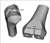
Figure 2
Medio-lateral width of the femoral implant at four points. Post, posterior; DP, distal posterior; DA, distal anterior; Ant, anterior.
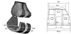
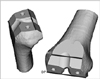
Figure 3
The aspect ratio was defined as the mediolateral width (ML) divided by the anteroposterior width (AP).

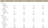
Figure 4
The width ratio was defined as the anterior (Ant) medio-lateral width divided by the distal posterior (DP) medio-lateral width.

References
1. Hitt K, Shurman JR 2nd, Greene K, et al. Anthropometric measurements of the human knee: correlation to the sizing of current knee arthroplasty systems. J Bone Joint Surg Am. 2003; 85:Suppl 4. 115–122.
2. Cho WS, Moon HS, Park SS, Noh KM, Cha HI. The shape and size discrepancy between bone and prosthesis in total knee arthroplasty. J Korean Orthop Assoc. 1998; 33:1045–1055.
3. Seeman E. Periosteal bone formation: a neglected determinant of bone strength. N Engl J Med. 2003; 349:320–323.
4. Westrich GH, Laskin RS, Haas SB, Sculco TP. Resection specimen analysis of tibial coverage in total knee arthroplasty. Clin Orthop Relat Res. 1994; 309:163–175.
5. Kim JM, Kim SB, Kim JM, Lee DH, Lee BS, Bin SI. Results of gender-specific total knee arthroplasty: comparative study with traditional implant in female patients. Knee Surg Relat Res. 2015; 27:17–23.
6. Bae DK, Lee YH, Chung SW. The study of anatomical measurement of distal femur and compatibility of femoral prosthesis in total knee arthroplasty. J Korean Orthop Assoc. 2002; 37:204–210.
7. Bae DK, Park JY. The study of anatomical measurement of proximal tibia and fitness of tibial prosthesis in total knee arthroplasty. J Korean Orthop Assoc. 2000; 35:57–64.
8. Chung BJ, Kang JY, Kang YG, Kim SJ, Kim TK. Clinical implications of femoral anthropometrical features for total knee arthroplasty in Koreans. J Arthroplasty. 2015; 30:1220–1227.
9. Chin KR, Dalury DF, Zurakowski D, Scott RD. Intraoperative measurements of male and female distal femurs during primary total knee arthroplasty. J Knee Surg. 2002; 15:213–217.
10. Chin PL, Tey TT, Ibrahim MY, Chia SL, Yeo SJ, Lo NN. Intraoperative morphometric study of gender differences in Asian femurs. J Arthroplasty. 2011; 26:984–988.
11. Mahfouz MR, Merkl BC, Fatah EE, Booth R Jr, Argenson JN. Automatic methods for characterization of sexual dimorphism of adult femora: distal femur. Comput Methods Biomech Biomed Engin. 2007; 10:447–456.
12. Yue B, Varadarajan KM, Ai S, Tang T, Rubash HE, Li G. Differences of knee anthropometry between Chinese and white men and women. J Arthroplasty. 2011; 26:124–130.
13. Lim HC, Bae JH, Yoon JY, Kim SJ, Kim JG, Lee JM. Gender differences of the morphology of the distal femur and proximal tibia in a Korean population. Knee. 2013; 20:26–30.
14. Park HT, Choi SJ, Park JH, Ha JM. Anthropometric study of distal femur & compatibility of gender knee prosthesis in total knee replacement. J Korean Knee Soc. 2008; 20:169–174.
15. Moon JS, Yu KS, Ha CW. Anthropometric analyses of Korean female knees. J Korean Knee Soc. 2006; 18:121–126.
16. Seedhom BB, Longton EB, Wright V, Dowson D. Dimensions of the knee. Radiographic and autopsy study of sizes required by a knee prosthesis. Ann Rheum Dis. 1972; 31:54–58.
 XML Download