

 PDF
PDF ePub
ePub Citation
 Print
Print


Abstract
Angular deformity of extremities in children and adolescents with residual growth is not a rare condition in orthopaedics. Asymmetrical physeal suppression or guided growth method, one of the surgical techniques for correction of angular deformity, is a method of inducing plastic deformation by application of constant external force to a growing bone. Internal fixation devices used for asymmetrical physeal suppression include staples, transphyseal screws, and tension band plates, most representatively the 8-plate. Temporary hemiepiphysiodesis using staples is reported to show a success rate of 60% to 80%. Epiphysiodesis using transphyseal screws has several advantages over staples or 8-plates; smaller skin incision, shorter operation time, no postoperative splint or cast, faster return to daily life. Advantages of 8-plates over staples or transphyseal plates include a longer moment arm, which enables better correction of angular deformity and less suppression of the growth of the nearby normal growth plate. Asymmetrical physeal suppression is a simple and effective surgical method in correcting angular deformity of extremities of children and adolescents. Each of three internal fixation devices discussed in the current article has strengths and weaknesses and superiority in terms of angular correction power and complication rate, however further study is needed. Therefore, the most appropriate device should be selected according to the condition of each patient.
Figures and Tables
Figure 1
A 15-year-old boy with Ehlers-Danlos syndrome. He developed genu valgum of the left knee (A) which was over corrected using staples in consideration of expected rebound overgrowth (B).
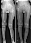
Figure 2
After temporary hemiepiphysiodesis with a staple, extrusion of the staple from the metaphysis occurred.
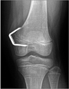
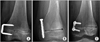
Figure 3
(A) An 11-year-old boy with cerebral palsy showed leg length discrepancy and right side genu valgum. Percutaneous epiphysiodesis using a transphyseal screw was performed for physeal growth suppression. (B) Correction of the leg length discrepancy and angular deformity was observed.
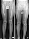
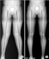
Figure 4
(A) Idiopathic bilateral genu valgum was observed in a 10-year-old girl. A tension band plate was used for asymmetrical physeal growth suppression. (B) Correction of the lower leg angular deformity was observed.

References
1. Villemure I, Stokes IA. Growth plate mechanics and mechanobiology. A survey of present understanding. J Biomech. 2009; 42:1793–1803.

2. Apte SS, Kenwright J. Physeal distraction and cell proliferation in the growth plate. J Bone Joint Surg Br. 1994; 76:837–843.
3. Stokes IA, Aronsson DD, Dimock AN, Cortright V, Beck S. Endochondral growth in growth plates of three species at two anatomical locations modulated by mechanical compression and tension. J Orthop Res. 2006; 24:1327–1334.
4. Bylski-Austrow DI, Wall EJ, Rupert MP, Roy DR, Crawford AH. Growth plate forces in the adolescent human knee: a radiographic and mechanical study of epiphyseal staples. J Pediatr Orthop. 2001; 21:817–823.
5. Frost HM. A chondral modeling theory. Calcif Tissue Int. 1979; 28:181–200.
6. Schroerlucke S, Bertrand S, Clapp J, Bundy J, Gregg FO. Failure of Orthofix eight-plate for the treatment of Blount disease. J Pediatr Orthop. 2009; 29:57–60.
7. Wiemann JM 4th, Tryon C, Szalay EA. Physeal stapling versus 8-plate hemiepiphysiodesis for guided correction of angular deformity about the knee. J Pediatr Orthop. 2009; 29:481–485.
8. Cho TJ, Choi IH, Chung CY, Yoo WJ, Park MS, Lee DY. Hemiepiphyseal stapling for angular deformity correction around the knee joint in children with multiple epiphyseal dysplasia. J Pediatr Orthop. 2009; 29:52–56.
9. Haas SL. Mechanical retardation of bone growth. J Bone Joint Surg Am. 1948; 30:506–512.
10. Blount WP, Clarke GR. Control of bone growth by epiphyseal stapling; a preliminary report. J Bone Joint Surg Am. 1949; 31:464–478.
11. Goyeneche RA, Primomo CE, Lambert N, Miscione H. Correction of bone angular deformities: experimental analysis of staples versus 8-plate. J Pediatr Orthop. 2009; 29:736–740.
12. Aykut US, Yazici M, Kandemir U, et al. The effect of temporary hemiepiphyseal stapling on the growth plate: a radiologic and immunohistochemical study in rabbits. J Pediatr Orthop. 2005; 25:336–341.
13. Stevens PM, MacWilliams B, Mohr RA. Gait analysis of stapling for genu valgum. J Pediatr Orthop. 2004; 24:70–74.
14. Stevens PM, Maguire M, Dales MD, Robins AJ. Physeal stapling for idiopathic genu valgum. J Pediatr Orthop. 1999; 19:645–649.
15. Mielke CH, Stevens PM. Hemiepiphyseal stapling for knee deformities in children younger than 10 years: a preliminary report. J Pediatr Orthop. 1996; 16:423–429.
16. Métaizeau JP, Wong-Chung J, Bertrand H, Pasquier P. Percutaneous epiphysiodesis using transphyseal screws (PETS). J Pediatr Orthop. 1998; 18:363–369.
17. Shin SJ, Cho TJ, Park MS, et al. Angular deformity correction by asymmetrical physeal suppression in growing children: stapling versus percutaneous transphyseal screw. J Pediatr Orthop. 2010; 30:588–593.
18. Ilharreborde B, Gaumetou E, Souchet P, et al. Efficacy and late complications of percutaneous epiphysiodesis with transphyseal screws. J Bone Joint Surg Br. 2012; 94:270–275.
19. De Brauwer V, Moens P. Temporary hemiepiphysiodesis for idiopathic genua valga in adolescents: percutaneous transphyseal screws (PETS) versus stapling. J Pediatr Orthop. 2008; 28:549–554.
20. Khoury JG, Tavares JO, McConnell S, Zeiders G, Sanders JO. Results of screw epiphysiodesis for the treatment of limb length discrepancy and angular deformity. J Pediatr Orthop. 2007; 27:623–628.
21. Stevens PM. Guided growth for angular correction: a preliminary series using a tension band plate. J Pediatr Orthop. 2007; 27:253–259.
22. Lee HJ, Oh CW, Song KS, Kyung HS, Min WK, Park BC. Guided growth with a noncannulated screw-plate system for angular deformity of the knee: a preliminary report. J Pediatr Orthop B. 2012; 21:339–347.
23. Stevens PM, Klatt JB. Guided growth for pathological physes: radiographic improvement during realignment. J Pediatr Orthop. 2008; 28:632–639.
24. Ballal MS, Bruce CE, Nayagam S. Correcting genu varum and genu valgum in children by guided growth: temporary hemiepiphysiodesis using tension band plates. J Bone Joint Surg Br. 2010; 92:273–276.
25. Burghardt RD, Herzenberg JE, Standard SC, Paley D. Temporary hemiepiphyseal arrest using a screw and plate device to treat knee and ankle deformities in children: a preliminary report. J Child Orthop. 2008; 2:187–197.
26. Stitgen A, Garrels K, Kobayashi H, Vanderby R, McCarthy JJ, Noonan KJ. Biomechanical comparison between 2 guidedgrowth constructs. J Pediatr Orthop. 2012; 32:206–209.
27. Ogden JA. The evaluation and treatment of partial physeal arrest. J Bone Joint Surg Am. 1987; 69:1297–1302.
28. Peterson HA. Partial growth plate arrest and its treatment. J Pediatr Orthop. 1984; 4:246–258.
29. Brockway A, Craig WA, Cockreli BR Jr. End-result study of sixty-two stapling operations. J Bone Joint Surg Am. 1954; 36:1063–1070.
30. Frantz CH. Epiphyseal stapling: a comprehensive review. Clin Orthop Relat Res. 1971; 77:149–157.
 XML Download