

 PDF
PDF ePub
ePub Citation
 Print
Print


The elbow is a common location for heterotopic ossification under certain circumstances. Trauma, neurologic injury, thermal burns, and surgery have all been associated with heterotopic ossification of the elbow.1) Postoperative heterotopic ossification of the elbow after surgery to treat acute trauma such as fracture/dislocation and tendon rupture has been well-documented.2) However, literature concerning heterotopic ossification after medial epicondylectomy in a non-acute setting is scarce. We report two cases of heterotopic ossification that occurred following medial epicondylectomies; One patient had medial epicondylitis with possible coincidental ulnar neuritis and another had cubital tunnel syndrome preoperatively.
CASE REPORT
1. Case 1
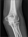
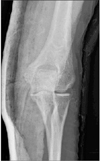
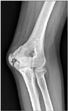
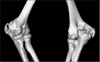
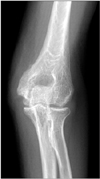
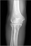
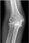
A 58-year-old female presented at our outpatient clinic with pain and limited range of motion of her left elbow. She had been diagnosed with medial epicondylitis with possible coincidental ulnar neuritis and had undergone medial epicondylectomy and debridement of the common flexor tendon origin of the symptomatic elbow, at another institution seven months prior to visiting to our clinic. Upon initial presentation, the patient's range of motion was limited to 135° flexion and 45° extension on the left elbow, compared to 150° flexion and 0° extension on the right side. Supination and pronation were not limited on either side. The patient complained of persistent pain at the medial aspect of the elbow and a tingling sense of her small finger. A review of her outside initial preoperative radiographs revealed only mild calcification at the medial epicondyle (Fig. 1). Immediate postoperative radiographs did not show any abnormal findings (Fig. 2). At the time of her visit to our clinic, seven months after the operation, radiographs revealed prominent heterotopic ossification of the elbow (Fig. 3). Computed tomography scans further revealed heterotopic ossification at the medial epicondyle, trochlea, posterior portion of the capitellum and olecranon (Fig. 4).
We performed surgery to remove heterotopic bone and relieve ankylosis. A posteromedial incision was made along the left elbow. The ulnar nerve was found to be entrapped within the heterotopic bone. Beginning from the medial supracondylar ridge, the posterior and anterior muscles were elevated, exposing the heterotopic bone. The heterotopic bone attached to the medial side of the olecranon was found to be blocking the elbow from full extension. We identified the ulnohumeral joint and carefully osteotomized and removed the heterotopic bone from the olecranon, and also removed the heterotopic bone on the posteromedial side of the medial epicondyle. After gentle manual arthrolysis, we achieved full range-of-motion at the elbow from 145° flexion to 10° hyperextension. Rotation of the forearm was full. Then we performed subcutaneous anterior transposition of the ulnar nerve.
After surgery, the patient was encouraged to perform gentle elbow range-of-motion exercises a few days postoperatively. Postoperative radiation therapy was not performed. Non-steroidal anti-inflammatory drugs were prescribed for heterotopic ossification prophylaxis. One year postoperatively, elbow range of motion was full with no evidence of heterotopic ossification on follow-up radiographs (Fig. 5).
2. Case 2
A 48-year-old male visited with limited range of motion of his right elbow. He had been diagnosed with cubital tunnel syndrome and had undergone cubital tunnel release and medial epicondylectomy at another institution one year prior to visiting to our clinic. The patient's range of motion was limited to 90° flexion and 0° extension, compared to 135° flexion and 0° extension on the left side. Supination and pronation were not limited on either side. His symptoms of ulnar nerve compression had improved, but the range of motion began to decrease postoperatively. His outside initial preoperative radiographs showed mild calcification at the medial epicondyle (Fig. 6). At the time we saw the patient, radiographs revealed heterotopic ossification at the medial and posterior sides of the elbow (Fig. 7). We considered that the heterotopic bone formed at the posterior oblique bundle of the medial collateral ligament was causing limitation of the elbow flexion. Although we recommended heterotopic bone removal and contracture release, the patient declined further operative treatment.
DISCUSSION
We described two cases of heterotopic ossification of the elbow following medial epicondylectomies for medial epicondylitis and for cubital tunnel syndrome.
Medial epicondylectomy is an effective treatment for treating cubital tunnel syndrome, and with debridement of pathologic tissue at the common flexor origin, it is recognized as one of the surgical options for medial epicondylitis.3) In the described cases, however, we speculate the medial epicondylectomy might be related with the occurrence of heterotopic ossification. It has been reported that the basic pathogenesis of heterotopic ossification involves transformation of primitive mesenchymal cells, present in the soft tissues of fascia, into osteogenic cells.4) An initiating event, such as trauma or underlying genetic disorders, induces local and/or systemic factors, including bone morphogenic proteins and prostaglandin-E2.5) This then leads to pathologic recruitment and induction/differentiation of osteoprogenitor cells, which in turn leads to proliferation of osteoblasts and a reduction in osteoclasts, resulting in heterotopic ossification. 5) Medial epicondylectomy in these patients may have provided the necessary triggers towards heterotopic ossification, such as the release of osteogenic precursor cells present in bone marrow.6)
In these patients, there were calcifications at the medial epicondyle preoperatively. Although the pathophysiology of calcific tendinopathy remains largely unknown, the presence of calcification may suggest a 'favorable environment' towards further mineralization. 7) Rather than being formed by precipitation of inorganic ions, calcific tendinopathy is now understood as an active cell-mediated process, in which resident progenitor cells with multi-differentiation potential, such as tendon-derived stem cells, may undergo chondral metaplasia and ectopic ossification by erroneous differentiation.7, 8) Therefore, pre-existing calcification, combined with further triggering, such as the release of osteogenic precursor cells by medial epicondylectomy in these cases, may be associated with heterotopic ossification.
However, there has been no report on heterotopic ossification following decortication or drilling of the bone, which is also often combined with debridement of tendons for the surgical treatment of medial epicondylitis.9) Furthermore, surgical removal of only calcifications in other anatomical areas such as the shoulder have yielded mostly favorable results, and there have been no reports of heterotopic ossification after such surgery.10) Nonetheless, the presented cases suggest that caution may be necessary when considering medial epicondylectomy in the presence of calcifications, since this combination may pose an increased risk of postoperative heterotopic ossification of the elbow.
 XML Download