

 PDF
PDF ePub
ePub Citation
 Print
Print


Tendon rupture is a rare complication of a distal radius fracture. Although the previously reported incidence of rupture of the extensor pollicis longus tendon after distal radius fractures varies from 0.07% to 0.88%, the incidence of rupture of flexor tendons following distal radius fractures is lesser than that of rupture of extensor tendons.1) Recently, a number of cases and mechanisms for chronic rupture of flexor tendons caused by attrition from volar plating have been reported.2) However, acute rupture of flexor tendons occurs less frequently than chronic rupture of flexor tendons. We experienced four cases of acute rupture of flexor tendons in association with distal radius fractures and have reported these cases along with literature review.
CASE REPORTS
1. Case 1
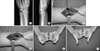
A 29-year-old right-handed male presented with painful swelling of his left wrist after rolling down the stairs. Physical examination revealed a loss of active flexion of the interphalangeal (IP) joint of the left thumb but other fingers did not have any limitation of active motion. The plain radiographs showed a dorsally displaced, comminuted intra-articular fracture with volar spike of distal radius and a fracture of the styloid process of the ulna (Fig. 1A, 1B).
The operation was performed through a volar radial approach. The rupture of flexor pollicis longus (FPL) tendon (Fig. 1C) and the laceration of pronator quadratus (PQ) muscle were identified at the level of the fracture of distal radius. The fracture was fixed with a volar locking plate and the ruptured FPL tendon was repaired with four-strand cruciate suture (Nylon 3-0) and epitendinous suture (Prolene 5-0) (Fig. 1D). After surgery, posterior thumb spica splint with extension block was applied for four weeks and gentle active motion of the IP joint of the left thumb was permitted immediately. Four weeks after the operation, a protective splint was applied for an additional two weeks. At the 13 months follow-up, the wrist was pain-free and a range of motion (ROM) was 80° palmar flexion, 80° dorsal extension, 80° supination and 80° pronation. And active motion of the IP joint of left thumb was not limited (Fig. 1E, 1F).
2. Case 2
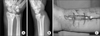
A 22-year-old right-handed male presented to the emergency department after a car accident. He had severe swelling and tenderness over his right wrist and an intact active motion of all fingers. The plain radiographs showed a dorsally displaced fracture with volar spike of distal radius and a fracture of the styloid process of ulna (Fig. 2A, 2B). Surgical treatment was planned because his fracture was unstable. Simultaneously, he also sustained a contralateral unstable fracture of distal radius.
Seven days after the trauma, open reduction and internal fixation through the volar radial approach was performed. The incision was made just radial to the flexor carpi radialis (FCR) tendon and the ruptured FCR tendon was identified immediately (Fig. 2C). Both ends of the ruptured FCR tendon were retracted in the opposite direction until about 3.5 cm. Also, the forearm fascia under the FCR tendon and PQ muscle was injured. Volar spike of the distal radius was visible through the torn PQ muscle. The fracture was reduced and fixed with a volar locking plate and the FCR tendon was repaired with cruciate suture and epitendinous suture. And the accompanying scapholunate dissociation was fixed with two Kirschner wires. After surgery, the sugar tong splint was applied for five days and it was replaced with a short arm thumb spica cast for another six weeks. Six weeks after surgery, the cast was taken off and the two Kirschner wires were removed. Then the patient underwent gradual mobilization with passive ROM exercise. At the 12 months follow-up, there was no wrist pain and the ROM was 80° palmar flexion, 80° dorsal extension, 80° supination and 80° pronation.
3. Case 3
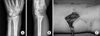
A 25-year-old right-handed male presented to the emergency department after a fall during mountain climbing. He had a limitation of motion of the left wrist due to pain but active motion of all fingers was intact. A laceration of about 1 cm in size was identified on the volar aspect of the wrist. And, tendon injury was not identified through the wound. Tingling or sensory changes in the fingers were not noted. Radiographs showed a fracture of the styloid process of the left distal radius and a fracture of the styloid process of the left ulna. The radiocarpal joint was dorsally dislocated and volar spike of the proximal margin of fractured radius was detected on radiographs (Fig. 3A, 3B). Simultaneously, he also sustained an ipsilateral clavicular fracture and a contralateral humeral shaft fracture.
The fracture of the radial styloid were exposed through a longitudinal incision over the radial styloid. It was fixed with a precontoured plate on the radial aspect of the radial styloid. An additional incision was made for the exploration of wound on the volar wrist. Intraoperatively, total rupture of the palmaris longus (PL) tendon was identified and repaired with cruciate suture (Fig. 3C). But, injury of other structures was not detected. Postoperatively, a short arm cast was applied for four weeks and then the patient underwent gradual mobilization with active assisted and gentle passive exercises. At the 16 months follow-up, the ROM of the left wrist was almost full and there were no other complications.
4. Case 4
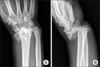
A 34-year-old right-handed male presented to the emergency department after a motorcycle accident. He sustained fractures of bilateral distal radius, fracture of the shaft of left radius and ulna and fracture of the shaft of left femur. The active flexion of right third finger was partially limited and the pain was aggravated by passive extension and flexion. Moreover, he complained of tingling sensation in his right palm in the dermatome of median nerve. Radiographs of the right wrist showed a dorsally displaced, comminuted intra-articular fracture with volar spike of distal radius and a fracture of the styloid process of ulna (Fig. 4).
The next day after the trauma, an operation was performed on right wrist. We planned an open reduction and internal fixation and exploration of the median nerve and flexor tendons. During carpal tunnel release, a partial rupture (>50%) of third flexor digitorum profundus (FDP) tendon was identified but an injury of median nerve was not found. The fracture was fixed with a volar locking plate and Kirschner wires and the ruptured FDP tendon was also repaired with four-strand cruciate suture (Nylon 3-0). Postoperatively, the sugar tong splint was applied for four weeks. After the splint was taken off, the wrist underwent gradual mobilization with active ROM exercise. At 18 months after the operation, the motion of the right third finger was intact and the wrist was 55° palmar flexion, 70° dorsal extension, 90° supination and 60° pronation.
DISCUSSION
Flexor tendon injuries following distal radius fractures are less common than extensor tendon injuries. Flexor tendons may be protected by the PQ and less tight enclosure over the distal radial aspect compared with extensor tendon.3) However, the incidence of rupture of the flexor tendons is unknown and has not been reported so far. Between March 2007 and December 2011, we experienced four cases with acute injury of flexor tendons including FCR, FPL, PL, and 3rd FDP in 474 patients with distal radius fractures that were treated with open reduction and internal fixation. And the incidence of acute rupture of the flexor tendons at our institute was 0.84%.
A literature search revealed one case of FCR rupture in association with distal radius fracture. DiMatteo and Wolf4) described a case of an acute FCR tendon rupture identified intraoperatively and found that it was caused by a volar spike of bone through the torn PQ. Because it is hard to detect the rupture of FCR tendon on physical examination of distal radius fractures, closed acute injury to the FCR tendon may be missed except the exploration using volar approach. In our case, torn PQ, bone spike of distal radius and two frayed ends of the FCR tendon with a 3.5 cm gap were intraoperatively identified with volar approach and this bone spike might have caused direct laceration of the FCR tendon.
The injury of FPL tendon in distal radius fracture may occur as acute or chronic rupture. Most of cases were chronic rupture after 4 weeks of distal radius fractures.5,6) McMaster,5) who first described a rupture of FPL tendon after distal radius fracture, suggested that two different pathological processes were responsible for this occurrence. First process involved a direct partial laceration of FPL tendon by the bone spur at the time of fracture and the laceration was not completely healed during the period of immobilization, thereby resulting in a weak spot and later a strong exertion on the tendon caused the tendon rupture. Second process was a local necrosis due to a decrease of blood supply caused by rupture or obstruction of the blood supply with additional pressure due to the hematoma or callus. Only two cases were reported for acute rupture of the FPL tendon that occurred within four weeks after distal radius fracture. However, these two cases had no immediate evidence of total rupture of the FPL tendon after fracture. Ruptures of the FPL tendon were indentified after 4 weeks in Wong and Pho's case7) and 3 days in Kim et al.'s case8), respectively. In our case, the patient could not flex the IP joint of his thumb on initial examination and the rupture of FPL tendon was intraoperatively confirmed on the next day.
Although the rupture of PL tendon into the palmaris fascia as a cause of an acute carpal tunnel syndrome has been reported, there is no report of a case of rupture of the PL tendon following distal radius fracture.9) In this case, there was initially no evidence of rupture of PL tendon. However, the rupture was found by the exploration of laceration during operation for internal fixation.
The rupture of FDP or superficialis tendons has been reported more frequently than that of FPL tendon. However, most of these reports were chronic rupture of these tendons. Only Southmayd et al.10) reported a case of rupture of second FDP tendon that occurred soon after a distal radius fracture. In our case, the rupture of third FDP tendon was preoperatively not identified. The injury of third FDP tendon rupture was intraoperatively identified and it was repaired. Most of distal radius fractures in our clinics have been immobilized for 4 weeks. The postoperative immobilized period for distal radius fractures was not changed because of the rupture of flexor tendon. The repair of FPL or FDP injury was protected by extension block splint and active motion of injured finger was permitted.
In our cases, all acute ruptures occurred in young adults by high-energy injuries. Although this ruptures can caused by low-energy injuries, most of reported cases were related with high-energy injuries.4,7,8,10) However, we think that it is hard to confirm the effect of the mechanism of injury because acute ruptures of flexor tendons following distal radius fracture is very rare. Rather, all distal radius fractures of this study showed a severe displaced and volar-spiked pattern that might be likely to cause acute ruptures of flexor tendon. And acute pain by the fracture may interrupt active motion of ipsilateral fingers. Therefore, the authors think that the careful examination have to be performed in order to identify acute ruptures of flexor tendon that may complicates in distal radius fractures.
 XML Download