

 PDF
PDF ePub
ePub Citation
 Print
Print


To prevent complications from delayed rehabilitation and to promote early recovery of joint function after a total knee arthroplasty (TKA), various measures have been suggested for treatment of postoperative knee stiffness.1) Manipulation under anesthesia is an effective method for management of knee stiffness after TKA. Although manipulation has been reported to have a low complication rate, it can cause hemarthroses, wound dehiscence, supracondylar fractures, and even fatal pulmonary emboli.2,3) To our knowledge, however, compartment syndrome of the thigh following manipulation for treatment of knee stiffness after TKA has not been previously reported.
Compartment syndrome is a rare but potentially devastating complication following TKA. If not detected early and if immediate surgical decompression is performed, permanent damage can to the skeletal muscle and neurovascular structures can occur.
The purpose of the report is to present a case of compartment syndrome of the thigh after manipulation in a case of knee stiffness following TKA.
CASE REPORT
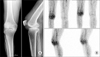
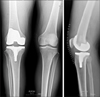
Here, we present the case of a 67-year-old man who underwent a right TKA for osteoarthritis. Three months earlier at a local hospital, he received a bee-sting therapy and an intra-articular steroid injection in his right knee where he later complained of painful swelling, localized heating sensation, and redness. With the impression of septic arthritis, he underwent arthroscopic synovectomy twice and intravenous antibiotic therapy. It was apparent that pus was present in the turbid synovial fluid obtained from an arthroscopic surgery. Further medical history was unremarkable and no oral anticoagulant was administered prior to surgery. Physical examination at the first visit revealed moderate swelling of the right knee joint and limited knee joint range of motion (ROM, 20°-80°; extension lag, 20°), while plain radiography showed degenerative osteoarthritis with a varus deformity (Fig. 1A). Blood serum examination revealed a white blood cell (WBC) count of 7,960 (4.8-10.8)×103/µl. The erythrocyte sedimentation rate and C-reactive protein level were elevated to 40 (normal range: below 9) mm/h and 8.0 (normal range: below 5) mg/L. Analysis of fluid aspirated from the affected joint revealed a WBC count of 3,000/mm3 with 60% neutrophils, glucose of 105 mg/dl and protein of 328 mg/dl. However, in both 2- and 4-hours delayed inflammation scan images (Tc-99m hexamethylpropyleneamine oxime WBC), WBC accumulation within the right knee soft tissue was observed (Fig. 1B). Thus, we suspected infectious arthritis of the knee and performed a frozen biopsy of several inner regions (anterior fat pad, superior, posterior, medial and lateral synovium) of the right knee joint in the operating room. All regions showed chronic inflammatory tissue with no polymorphonuclear leukocytes. We performed an extensive synovectomy to remove the hypertrophied synovium followed by an primary TKA (Genesis II Total Knee Replacement System, PS type; Smith & Nephew, Memphis, TN, USA) (Fig. 2). A sufficient soft tissue release was performed for treatment of soft tissue contracture. A Barovac (Sewoon, Seoul, Korea) was inserted after deflating tourniquet and adequate bleeding control of active bleeding sites. The patient has not taken any anti-thrombotic medication. The Barovac was removed three days later. The patient started performing straight-leg raises and quadriceps isometric exercises immediately after the surgery as well as the ROM exercise using continuous passive motion on the second postoperative day. However, because of pain, he had difficulty performing the scheduled ROM exercises. Thus, a fentanyl transdermal patch was added to the routine postoperative pain regimen, and the intravenous patient-controlled analgesia was continued.
On the day 28 postoperative, the ROM in the right knee was 0° to 60°. To restore the ROM of the involved knee, we performed a manipulation under anesthesia. Gentle controlled pressure was applied on the tibia close to the joint line to mitigate the risk of tibial fracture. After the manipulation, ROM of 0° to 100° was obtained.
On the second post-manipulation day, however, pain on the patient's distal thigh and knee was aggravated. Swelling and tightness of the anterolateral aspect of the right thigh became evident, and the motion of the hip and knee became limited due to severe pain.
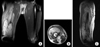
A radiographic examination of his extremities did not reveal any fracture or bony change. Magnetic resonance imaging (MRI) of the knee revealed a 10×8×10 cm sized fluid collection spreading over the mid-thigh vastus intermedius, vastus medialis and vastus tlearalis with the connection of the right knee joint (Fig. 3).
Under the suspicion of compartment syndrome of the right thigh, the pressure of each compartment was measured using the technique introduced by Whitesides et al. The mean pressures of the anterolateral and medial area were 55 and 50 mmHg, respectively, whereas the mean pressure of the posterior compartment was 25 mmHg. The Whitesides method, however, did not yield accurate or consistent results,4,5) so an arterial line manometer5) was used in the operating room to confirm the values. The mean pressures of antero-lateral and medial area were measured 45 and 41 mmHg. The patient underwent an emergency fasciotomy through a 10-cm longitudinal incision in the lateral side of the thigh. Inspection of the operation field found that a tear of soft tissue adherent to the distal thigh and hematoma of approximately 500 ml between the vastus lateralis and vastus intermedius. Because no active bleeding was observed, we performed decompression of the site simply by removing the hematoma.
The patient was discharged after four weeks. He has had no further complaints after the surgery, and the ROM in his right knee was 0° to 110° at the 36-month follow-up. The knee function score from the hospital for special surgery was 90, the Knee Society score was 90/90 (knee/function), and the Western Ontario and McMaster Universities arthritis index was 8.
DISCUSSION
Stiffness, one of the most frequent complications of TKA, may lead to suboptimal functional outcomes and decreased patient satiscfation. Despite improvements in prosthetic design and instrumentation in modern TKA, various studies have reported that postoperative knee stiffness occurs in 1.3% to 25% of patients.6) Various factors may lead to stiffness after TKA, including preoperative range of motion, prosthetic geometry, surgical technique, postoperative rehabilitation, and wound-healing problems.6,7) Occult infection is another consideration in the management of post-TKA stiffness. A low-grade infection may cause an underlying inflammation and pain, which then limits the patient's ability to participate in therapy.8)
Manipulation under anesthesia with or without arthroscopic adhesiolysis is the first tool for managing postoperative TKA stiffness and can effectively handle limited flexion. However, although manipulation has been reported to have a low complication rate, it can cause periprosthetic fractures, wound dehiscence, patellar tendon ruptures, and pulmonary emboli.2,3) To the best of our knowledge, compartment syndrome of the thigh following the manipulation for treatment of knee stiffness after TKA has not been previously reported. Total incidence of compartment syndrome after TKA is low, however, if it occurs, the common site is usually the calf. This is likely because the compartments surrounding the joint are mostly not involved in the procedure and the bleeding occurs in a joint cavity or leaks out through the wound.9)
In the literature, we found only four cases of compartment syndrome of the thigh after TKA that resulted in fasciotomies. Possible causes included a history of femoropopliteal bypass and iatrogenic coagulopathy due to heparinization. Other possible etiologic factors include extensive soft tissue dissection, an unnecessarily high tourniquet pressure, venous insufficiency through thrombus formation, and direct major vessel injury with subsequent reperfusion.10,11)
Our patient underwent two times of arthroscopic synovectomy to treat septic arthritis 3 months earlier in the same leg with underlying degenerative arthritis. In addition, the periarticular soft tsiuse showed increased vascularity induced by inflammation. Consequently, we performed a TKA simultaneously with an extensive tissue synovectomy to address the inflamed tissue. This aggressive handling of the soft tissue may have been a cause of the high-intensity postoperative pain, which impeded a smooth rehabilitation and required a longer recovery time for soft tissue healing.
Regarding the timing of manipulation, Yercan et al.6) stated that early manipulation provided better ROM gains than later manipulation, whereas Cates and Schmidt12) reported that manipulation was most effective within eight weeks, with full extension and at least 90° of flexion prior to manipulation. It is usually performed 6 to 12 weeks postoperatively. We performed the manipulation in the fourth week and did not exclude the possibility that this was too early. In this case, however, we judged it important to perform an early manipulation under anesthesia because of the high risk of knee stiffness resulting from multiple factors, such as the limited pre-arthroplasty range of articular motion, periarticular soft tissue inflammation, high-intensity postoperative pain, and prolonged rehabilitation period despite adequate pain management. Nevertheless, this resulted in recurrent damage to the tissue adherent around the scar tissue in a state of insufficient soft tissue healing, leading to the development of a large hematoma. This may have incurred superior expansion into the compartment of the thigh and subsequently caused compartment syndrome.
MRI of the knee revealed a fluid collection, measuring 10×8×10 cm size, spreading over the mid-thigh vastus intermedius, vastus medialis and vastus lateralis with the connection of the right knee joint. The patient underwent an emergency fasciotomy through a 10-cm longitudinal incision in the lateral side of the thigh, which revealed the injury to the soft tissue that was severely adhered to the perihematomal region but did not reveal any significant bleeding source.
A diagnosis of compartment syndrome requires a high index of suspicion and the use of adjunct modalities to ensure prompt treatment. The six Ps - pain, pallor, paresthesia, pulselessness, paralysis, and poikilothermia - may be found during patient presentation
When compartment syndrome is suspected following TKA, compartment pressure measurements using a Stryker needle unit will assist in diagnostic confirmation. The needle is inserted directly into the compartment in question, and a pressure >30 mmHg indicates an emergency requiring prompt treatment with a fasciotomy. In our case, pressure measurements of all three compartments were performed before and after the fasciotomy. The latter measurements showed reduction to normal levels.
Compartment syndrome of the thigh following manipulation for treatment of knee stiffness after TKA is a rare but potentially devastating complication. When performing manipulation to treat knee stiffness after TKA, clinicians should be aware of this complication in order to prevent prolonged ischemia and permanent damage. In this report, we identified manipulation for control of knee stiffness concomitant with periarticular inflammation as a possible risk factor. Due to the high risk of severe scar tissue adhesion and increased vascularity, special care should be taken in cases in which an extensive synovectomy is performed simultaneously with TKA.
 XML Download