

 PDF
PDF ePub
ePub Citation
 Print
Print


For many years patients with medial compartment osteoarthritis and varus deformity have been successfully treated with high tibial osteotomy (HTO).1) Recently, opening-wedge osteotomy has been mentioned to be more advantageous than closing wedge technique.2) Open wedge HTO has been more popular and with good results due the development of locking plate.3)
However the disadvantage of open wedge HTO is the empty space on the proximal tibia which is needed to be filled in with a spacer to gain union (autograft, allograft, etc.).4) Recently, hydroxyapatite (HAp) and beta-tricalcium phosphate (TCP) blocks are being used to fill the vacant space and through studies, TCP ceramics were shown to have superior absorption rate and osteoconductivity.1)
The authors report two patients who had a non-union at the osteotomy site after the HTO using a TCP block.
CASE REPORT
1. Case 1
A 53-year-old woman was admitted due to left knee pain. The range of knee motion was 0° to 115° and medial joint line tenderness was positive.
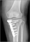
The patient received arthroscopic partial meniscectomy for medial meniscus posterior root tear, and arthroscopic chondroplasty for medial femoral condyle chondral lesion. Correction of 11° was made in order to move weight bearing line to 62% lateral of the proximal tibia. To achieve 11° correction, 13 mm is required, which is the osteotomy site length (6.2 cm) multiplied by the tangent 11°. Opening-wegde HTO with TOMOFIX (Synthes, Oberdorf, Switzerland), and 13 mm TCP block (Synthes) insertion was performed (Fig. 1).
For the initial 6 weeks, partial weight bearing with crutch was permitted and range of motion exercise was performed. After 6 weeks, tolerable weight bearing was allowed and muscle strengthening exercise was done.
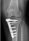
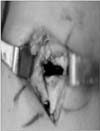
At 12 months after surgery, on the plain radiograph, nonunion of the wedge was seen (Fig. 2). Implant removal was performed. Most of the TCP blocks were nonabsorbed. Remaining TCP was removed, and then a 3×2 cm vacant space was observed intraoperatively (Fig. 3). The space due to nonunion was untouched because the good union state of the other portions.
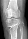
At 3 months after implant removal and anteroposterior radiograph also showed a radiolucent space with a well maintained correction angle (Fig. 4).
2. Case 2
A 55-year-old woman was admitted due to right knee pain. The range of knee motion was 14° to 98° and medial joint line tenderness was positive. The patient had a varus angle of 8.1° and Kellgren-Lawrence grade 2 OA change.
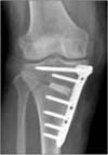
The patient received arthroscopic partial menisectomy for medial meniscus tear, arthroscopic microfracture for medial femoral condyle chondral lesion, and HTO with TOMOFIX (Synthes), TCP block (10 mm) for genu varum (Fig. 5).
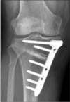
At 12 months, radiolucency was seen around the spacer on the plain radiograph (Fig. 6). Implant removal was done and TCP from the nonunion site was removed. The space due to nonunion was untouched because the good union state of the other portions
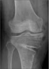
At 3 months after implant removal, standing anteroposterior radiograph also showed a radiolucent space and a well maintained angle (Fig. 7).
DISCUSSION
HTO is a procedure which is performed in patients with medial compartment degenerative disease, osteoarthritis, and varus deformity which delay the time until TKA is needed. Many studies have shown good results of HTO due to the development of TOMOFIX (Synthes).2456) Open wedge HTO showed better results than closed wedge HTO.2)
The advantages of open wedge HTO includes less risk of peroneal nerve injury and less instability of lateral knee ligament, facilitated technique, maintenance of bone stock, and better correction achievement.1) It has brought satisfactory clinical results for patients with medial unicompartmental osteoarthritis.7)
The disadvantages include the need for filling in the free space after osteotomy (autogenous bone graft causing donor site pain), delayed union and nonunion.289) TCP and HAp grafts are used to help with the union process and many studies have shown the efficacy of them both with TCP being a better choice.6) Onodera et al.6) reported that TCP had a higher osteoconductivity and greater absorbability than HAp spacer.
Saragaglia et al.10) reported the outcome of opening wedge HTO augmented with a Biosorb® (TCP) wedge in 124 patients with a mean of 10 years follow-up. There were only 7 knees with delayed union which required deferring weight-bearing. They reported the Biosorb® wedge to be completely integrated in all cases.
Present study demonstrated the 2 cases out of 38 cases (5.2%) from March 2012 to March 2014 of nonunion with the use of TCP block. Both patients are nonsmokers, and did not have a history of steroid use. Other complications, excluding nonunion of two cases, did not occur.
In both cases, correction angle was maintained at 3 months after implant removal. The reason why the angle was well maintained was because the area without TCP block showed good union.
The authors of this study reports that the use of TCP block in HTO procedure may interfere with the process of union. Therefore caution is thought to be needed when choosing the spacers when performing HTO.
 XML Download