

 PDF
PDF ePub
ePub Citation
 Print
Print


Abstract
Atlas fracture accounts for 1% to 3% of all spinal column injuries and 10% of cervical spine fractures, and is most frequently caused by motor vehicle accidents and falls. Only a few cases involving complications after surgical treatment have been reported. We present a case of anterior atlas arch stress fracture accompanied by worsening neurologic symptoms following atlas posterior arch resection for cervical myelopathy with retro-odontoid pseudotumor.
Figures and Tables
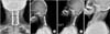
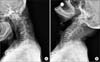 | Figure 1Lateral cervical radiographs showing severe degenerative spondylosis, kyphosis and instability at C1-C2. The atlanto-dental interval is 5 mm at flexion (A) and reduced at extension (B).
|
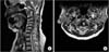 | Figure 2The retro-odontoid pseudotumor and intramedullary hyper-intense lesion of C1 and C2 were observed in sagittal (A) and axial (B) images on T2-weighted magnetic resonance imaging.
|
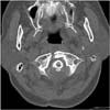 | Figure 3Computed tomography scans showing that C1 posterior arch resection was accomplished and that C1 anterior fracture occurred.
|
References
1. Isono M, Ishii K, Kamida T, Fujiki M, Goda M, Kobayashi H. Retro-odontoid soft tissue mass associated with atlantoaxial subluxation in an elderly patient: a case report. Surg Neurol. 2001; 55:223–227.
2. Yamaguchi I, Shibuya S, Arima N, Oka S, Kanda Y, Yamamoto T. Remarkable reduction or disappearance of retroodontoid pseudotumors after occipitocervical fusion. Report of three cases. J Neurosurg Spine. 2006; 5:156–160.
3. Suetsuna F, Narita H, Ono A, Ohishi H. Regression of retroodontoid pseudotumors following C-1 laminoplasty. Report of three cases. J Neurosurg Spine. 2006; 5:455–460.
4. O'Shaughnessy BA, Salehi SA, Ali S, Liu JC. Anterior atlas fracture following suboccipital decompression for Chiari I malformation. Report of two cases. J Neurosurg Spine. 2004; 1:137–140.
5. Hirano Y, Sugawara A, Mizuno J, Takeda M, Watanabe K, Ogasawara K. Spontaneous C1 anterior arch fracture as a postoperative complication of foramen magnum decompression for Chiari malformation type 1. Surg Neurol Int. 2011; 2:138.
6. Jun BY, Yoon KJ, Crockard A. Retro-odontoid pseudotumor in diffuse idiopathic skeletal hyperostosis. Spine (Phila Pa 1976). 2002; 27:E266–E270.
7. Yoshida K, Hanyu T, Takahashi HE. Progression of rheumatoid arthritis of the cervical spine: radiographic and clinical evaluation. J Orthop Sci. 1999; 4:399–406.
8. Cihanek M, Fuentès S, Metellus P, Pech-Gourg G, Dufour H, Grisoli F. Disappearance of retro-odontoid pseudotumor after C1-C2 transarticular fixation screw. Neurochirurgie. 2008; 54:32–36.
9. Chikuda H, Seichi A, Takeshita K, et al. Radiographic analysis of the cervical spine in patients with retro-odontoid pseudotumors. Spine (Phila Pa 1976). 2009; 34:E110–E114.
10. Kakutani K, Doita M, Yoshikawa M, et al. C1 laminectomy for retro-odontoid pseudotumor without atlantoaxial subluxation: review of seven consecutive cases. Eur Spine J. 2013; 22:1119–1126.
 XML Download