

 PDF
PDF ePub
ePub Citation
 Print
Print


Abstract
Purpose
We performed clinical and radiological evaluation of surgical outcomes of congenital vertical talus.
Materials and Methods
Fifteen surgically treated feet in 9 patients (6 bilateral and 3 unilateral) which were followed-up for at least 2 years were included. Mean patient age at the time of surgery was 10.9 months. The surgical technique was a one-stage correction using the Kumar technique with a Cincinnati skin incision. In 7 feet we also transferred half of the tibialis anterior to the talar neck (the Grice technique). Radiologic parameters (talo-calcaneal angle, talo-first metatarsal angle, tibio-talar angle, tibio-calcaneal angle) were analyzed pre- and postoperatively and at the last follow-up, and clinical outcomes by the Laaveg-Ponseti score.
Results
Talus orientation was improved in all patients. All radiologic parameters showed statistically significant improvement by the last follow-up. The mean Laaveg-Ponseti score at the last follow-up was 16 for patient satisfaction, 16 for function, and 24 for pain. There was no recurrence, however one case of talar neck fracture occurred during the tibialis anterior transfer.
Figures and Tables
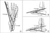 | Figure 1Radiologic parameters measured in anteroposterior (A) and lateral (B, C) views: talo-calcaneal angle (T-C angle: the angle between the mid-talar and mid-calcaneal axes); talo-first metatarsal angle (T-M angle: the angle between the mid-talar and mid-first metatarsal axes); tibio-talar angle (Ti-T angle: the angle between the mid-tibia and mid-talar axes); tibio-calcaneal angle (Ti-C angle: the angle between the mid-tibia and mid-calcaneal axes).
|
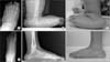 | Figure 2Anteroposterior (A) and lateral (B) radiographs of a 10-month-old patient (case 3) with left congenital vertical talus. (C) Photograph showing a rocker-bottom deformity. (D, E) Radiographs taken 7 years after surgery (when the patient was 8 years old) showing correction of the vertically oriented talus. (F) Photograph taken at the last follow-up shows satisfactory correction of the deformity.
|


References
1. Becker-Andersen H, Reimann I. Congenital vertical talus. Reevaluation of early manipulative treatment. Acta Orthop Scand. 1974; 45:130–144.
2. Dobbs MB, Purcell DB, Nunley R, Morcuende JA. Early results of a new method of treatment for idiopathic congenital vertical talus. J Bone Joint Surg Am. 2006; 88:1192–1200.
3. Alaee F, Boehm S, Dobbs MB. A new approach to the treatment of congenital vertical talus. J Child Orthop. 2007; 1:165–174.
4. Choi IH, Chung CY, Cho TJ, Yoo WJ, Park MS. Duk Yong Lee's pediatric orthopaedics. 4th ed. Seoul: Koonja;2014. p. 517–521.
5. Kumar SJ, Cowell HR, Ramsey PL. Vertical and oblique talus. Instr Course Lect. 1982; 31:235–251.
6. Colton CL. The surgical management of congenital vertical talus. J Bone Joint Surg Br. 1973; 55:566–574.
7. Grice DS. An extra-articular arthrodesis of the subastragalar joint for correction of paralytic flat feet in children. J Bone Joint Surg Am. 1952; 34:927–940.
8. Coleman SS, Stelling FH 3rd, Jarrett J. Pathomechanics and treatment of congenital vertical talus. Clin Orthop Relat Res. 1970; 70:62–72.
9. Lakshmanan P, Phillips SJ, Thomas RH, O'Doherty DP. Partial wound closure of the Cincinnati incision in clubfoot correction. Eur J Orthop Surg Traumatol. 2005; 15:28–31.
10. Grice DS. The role of subtalar fusion in the treatment of valgus deformities of the feet. Instr Course Lect. 1959; 16:127–150.
11. Laaveg SJ, Ponseti IV. Long-term results of treatment of congenital club foot. J Bone Joint Surg Am. 1980; 62:23–31.
12. Lamy L, Weissman L. Congenital convex pes valgus. J Bone Joint Surg Am. 1939; 21:79–91.
13. Osmond-Clarke H. Congenital vertical talus. J Bone Joint Surg Br. 1956; 38:334–341.
14. Lloyd-Roberts GC, Spence AJ. Congenital vertical talus. J Bone Joint Surg Br. 1958; 40:33–41.
15. Eyre-Brook AL. Congenital vertical talus. J Bone Joint Surg Br. 1967; 49:618–627.
16. Dodge LD, Ashley RK, Gilbert RJ. Treatment of the congenital vertical talus: a retrospective review of 36 feet with long-term follow-up. Foot Ankle. 1987; 7:326–332.
17. Sharrard WJ, Grosfield I. The management of deformity and paralysis of the foot in myelomeningocele. J Bone Joint Surg Br. 1968; 50:456–465.
18. Wirth T, Schuler P, Griss P. Early surgical treatment for congenital vertical talus. Arch Orthop Trauma Surg. 1994; 113:248–253.
19. Harrold AJ. Congenital vertical talus in infancy. J Bone Joint Surg Br. 1967; 49:634–643.
20. Drennan JC, Sharrard WJ. The pathological anatomy of convex pes valgus. J Bone Joint Surg Br. 1971; 53:455–461.
21. Sharrard WJ. Paralytic deformity in the lower limb. J Bone Joint Surg Br. 1967; 49:731–747.
22. Patterson WR, Fitz DA, Smith WS. The pathologic anatomy of congenital convex pes valgus. Post morten study of a newborn infant with bilateral involvement. J Bone Joint Surg Am. 1968; 50:458–466.
23. Vanderwilde R, Staheli LT, Chew DE, Malagon V. Measurements on radiographs of the foot in normal infants and children. J Bone Joint Surg Am. 1988; 70:407–415.
 XML Download