

 PDF
PDF ePub
ePub Citation
 Print
Print


Cavernous hemangioma, also known as cavernous angioma, cavernous malformation or cavernoma is a developmental hamartomatous malformation. Extraosseous, extradural cavernous hemangiomas are rare and are usually presented as chronic progressive spinal cord syndrome, radiculopathy or a mere local back pain.1,2,3) An acute onset of symptoms is uncommon and if present, it is mostly caused by hemorrhage.1,2,3) We report a case a unique case of cavernous hemangioma showing acute symptoms without hemorrhage as well as chronic symptoms. The rarity of our case stresses a need to consider cavernous hemangioma as one of different diagnosis of extradural thoracic compressive myelopathy.
CASE REPORT
A 31-year-old male presented with a severe back pain and weakness of both lower extremities. He recalled having similar symptoms five and two years ago, working in a sitting position for a long time, but the pain relieved spontaneously when he rested in a lying position. He had mild chronic back pain but it was tolerable to a degree that he did not have to seek medical care. He was an office worker and had no problems in executing daily life activities. He had no past medical history, no history of trauma, fever, deformity, weight loss, bowel, or bladder dysfunction. The patient recalled that acute, excruciating pain suddenly developed when he was sleeping after a long day of work in a sitting position. When the patient visited the emergency room, all of both lower extremities motor grades were nearly grade 0, and there was a sensory impairment to all modalities below the twelfth thoracic dermatome. Hematological and biochemical investigations were within normal limit.
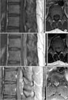
Computed tomography of the spine showed an extradural mass at the T11-T12 vertebral level without bony destruction. Magnetic resonance imaging (MRI) of the spine revealed an extradural lesion, opposite the T11-T12 vertebral bodies. The lesion was homogenously isointense on T1 and T2 weighted images and showed homogenous contrast enhancement on T1 weighted images (Fig. 1). The lesion was located dorsally and had compressed the thoracic cord ventrally. The spinal cord at the level was displaced ventrally, and there was no signal change of the cord.
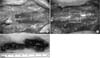
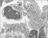
The patient underwent a T11-T12 laminectomy. The mass was blackish red in color, soft and friable. It was located epidurally and dorsal to the thecal sac. The lesion was peeled off the surface of the dura easily with mild adhesion (Fig. 2). There was no tear and subdural or intradural lesion on dura. Multiple proliferation of dilated vessels with flattened endothelium and blood cells inside and fibrofatty connective septae between vessels were noted in the histopathological examination, findings consistent with cavernous hemangioma (Fig. 3).
The patient was discharged five days after surgery. Postoperatively, both lower extremities motor grade recovered to grade V and all somatosensory system returned to nearly normal.
DISCUSSION
Cavernous hemangioma is a benign vascular malformation which can occur anywhere in the central nervous system.2,4) Pure epidural cavernous hemangioma is only rarely noted with about 100 cases reported in the literature thus far.5) Common clinical presentation is a progressive myelopathy or radiculopathy. Rarely, there may be acute symptoms with a significant neurologic deficit, and it can be explained by a sudden increase in volume of the lesion, mainly due to hemorrhage, or thrombosis of the draining veins.1,3) In a study reported by Aoyagi et al.,1) hemorrhage was absent only in one of five patients with acute myelopathy. However, in our case, the patient had no prodromal symptoms except for chronic back pain that was tolerable without any medication. Yet when he was sitting for a long time, a sudden onset of severe back pain with a rapid progress of paraparesis occurred. During surgery there was no definite hematoma or active bleeding to pinpoint hemorrhage as a cause of acute myelopathy.
Cavernous hemangiomas usually are isointense on T1 and isointense to hyperintense on T2 weighted images in MRI. Contrast enhancement frequently occurs.6) In our patient the lesion was isointense in T1 and T2 weighted images, and homogenous contrast enhancement was seen. Differential diagnoses are schwanoma, meningioma and metastatic tumor. Surgical total excision of the lesion is treatment of choice and it is possible for most of the lesions located posteriorly.1,2,7) When total excision is not possible adjuvant radiotherapy is advised.8) In our patient the lesion located posteriorly only and had no foraminal involvement so it was easily removed. With the sudden onset of severe neurologic deficit, a perfect result is doubtful and may result in disability or mortality.1,2,9) Our patient recovered completely after immediate surgery and shows well-maintained clinical results two months into follow-up. However, a longer follow-up period may be necessary to detect secondary lesions or signs of recurrence.
 XML Download