

 PDF
PDF ePub
ePub Citation
 Print
Print


Cuff tear arthropathy represents a broad spectrum of pathology in which, at least to some degree, three critical features are present: rotator cuff insufficiency, degenerative changes of the glenohumeral joint, and superior migration of the humeral head. Other characteristics that may be seen are humeral head collapse, erosive changes to the superior glenoid or acromion, and subdeltoid effusion. The reverse total shoulder arthroplasty has recently received attention for the treatment of rotator cuff tear arthropathy. However, many complications of reverse total shoulder arthroplasty have also been reported, and infection is one of the major complications.1,2,3) Infection rates after reverse total shoulder arthroplasty have been reported at approximately 1% to 10%.4,5,6,7,8) Among these cases, infection caused by Mycobacterium tuberculosis has not been reported. However, we are reporting the case of an approximately a 77-year-old female patient who underwentreverse total shoulder arthroplasty with surgical site infection caused by M. tuberculosis. The patient granted permission for her case to be reported.
CASE REPORT
A 77-year-old woman visited our hospital because of heat, pain and pus discharge at the surgical site. She had underwent reverse total shoulder arthroplasty for a rotator cuff tear arthropathy two years and three months ago. And she had underwent irrigation and debridement two times due to infectious signs. But no symptoms had been improved, so she had been refered to our hospital.
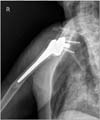
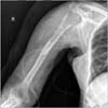
The medical history of the patient indicates that she was diagnosed with diabetes mellitus three years ago, and she was on medication for angina pectoris. There was no past history of immunocompromising disease. Her chief complaint was pus discharge, pain and swelling at the surgical site, 8 months after a reverse total shoulder arthroplasty. Physical examination revealed a drainage opening at the front of the right shoulder, which was in the middle area of the previous skin incision site and measured about 1 cm in diameter. The drained fluid was a dark yellowish color with relatively light viscosity. At that time we performed a Gram stain and culture, and analyzed for C-reactive protein (CRP) and erythrocyte sedimentation rate (ESR). CRP was 0.55 mg/L (0.5 mg/L, normal range) and ESR was 17 mm/h (0-30 mm/h, normal range). The results were within normal range because the patient was referred to our hospital after antibiotic treatment. In two Gram stain and bacterial culture tests, the first Gram stain isolated the Gram-positive cocci and the bacterial culture test cultured alpha-hemolytic streptococcus. At that time, the acid fast bacilli (AFB) stain and culture for M. tuberculosis test was not performed. At hospital admission, loosening of the prosthesis was not observed in a simple X-ray (Fig. 1).
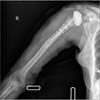
The first surgery was for removal of the previous prosthesis. Extensive
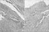
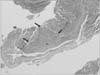
marginal resection was performed to remove infection and tissue necrosis. Irrigation with a large amount of normal saline and debridement was performed. All of the equipment was removed, including cement, and then the site was washed again in saline solution. Previous cement was removed with curved gauge. Vancomycin antibiotic powder 1 g was mixed with cement, which was inserted at the proximal humerus (Fig. 2). Intraoperative Gram stain and bacterial culture tests were performed, and all were negative. However, intraoperative biopsy findings showed acute inflammation with chronic granulomatous inflammation and focal foreign body reaction (Fig. 3). Heating and swelling after the operation had decreased by the 16th day, then the CRP was 0.76 mg/L and ESR was 62 mm/h. After an intravenous antibiotic was injected more 4 weeks, she was discharged and followed-up with oral antibiotics in the outpatient clinic.
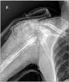
After six weeks in the outpatient clinic, heating, swelling and pain recurred again. At the time, CRP and ESR was 7.3 mg/L and 78 mm/h, respectively. So, we started intravenous first-generation cephalosporin. As we suspected, there was a recurrence of infection, so an incision and drainage was carried out. The previous antibiotic-impregnated cement spacer was removed, and infected necrotic tissue was debrided extensively. Cement mixed with antibiotics was inserted to fill the empty space in the humeral shaft. We inserted a rubber drainage tube and, the incision site was sutured using wire (Fig. 4). The discharge was mildly viscous and relatively clear. Gram stain and bacterial culture was done during the operation, but did not find anything. Tests for tuberculosis (TB) were not performed. Fourteen days after surgery, sustained discharge, swelling and heating continued and, CRP and ESR showed 3 mg/L and 80 mm/h or more, respectively. Incision and marginal infected tissue resection was performed once again, and this time the cement mixed with antibiotics was not inserted (Fig. 5). After postoperative 17 days, no abnormalities were observed at the operative site. CRP and ESR results were normalized, which values were 0.43 mg/L and 31 mm/h, respectively. So the patient was discharged and oral antibiotics were prescribed.
After the first infection, investigation did not show evidence of a family history of TB infection, and the patient's respiratory symptoms were limited to wheezing due to cardiac originated asthma. When there were symptoms of recurrence, we performed the incision and drainage at the previous surgical site wound and took multiple intraoperative tissue samples. The AFB stain returned negative findings, but M. tuberculosis were cultured in two of the four samples, with less than five colonies. Intraoperative biopsy findings showed focal granulomatous reaction (Fig. 6). After obtaining these results, we did not hesitate to administer the rifampin, isoniazid, ethambutol and pyrazinamide therapy, as well as first generation cephalosporin treatment. And we didn't do any additional procedure for the operation site. After three weeks, the infection wound was closed, at which time the first generation cephalosporin was discontinued. However, the TB medication was administered orally for nine months. The outpatient follow-up result was satisfactory without recurrence. At the time of the latest follow-up, the degree of pain was checked 3 point in the VAS score. And forward elevation, abduction, external rotation at side and internal rotation at back of the shoulder were 40°, 30°, 20° and buttock, respectively.
DISCUSSION
In 2009, the World Health Organization (WHO) reported that the number of new-onset TB, including from both pulmonary and extra-pulmonary origins, was more than 5.8 million. The report published by the WHO showed that TB prevalence (including human immunodeficiency virus [HIV] infection) in the Republic of Korea was approximately 5.5%. The interaction of M. tuberculosis with the human host begins when droplet nuclei containing microorganism from infectious patients are inhaled. After infection, TB is not always expressed because of endogenous factors such as innate immunity, non-immunologic defense, and the degree of cell-mediated immune function. Typically, 10% of infected people express active TB. In this case, there was no family history of TB. There was a recurrent infectious surgical site wound, without a specific immunocompromised state, from the initial reverse total shoulder arthroplasty. Based on this fact, TB infection was likely from the initial event of infection, but we had overlooked the possibility.
The presence of sinus tract, suggests the presence of an atypical organism.9) As reported in other case studies involving joint infections caused by M. tuberculosis, another indication of a possible mycobacterial organism is the history of negative bacterial and fungal cultures.10) In our case, there was continued infection, and negative bacterial and fungal stains and cultures, so we suspected the infections were caused by M. tuberculosis. Further a biopsy during surgery was taken from various parts of the tissue, to increase the reliability of the results.10)
The M. tuberculosis infection can be often expressed in people with compromised immunity, mostly due to HIV infection or other immune deterioration in patients with cancer. In these people, TB infection should be ruled out at the beginning of treatment. However, this case of M. tuberculosis infection occurred in the shoulder after the initial operation. There was no evidence of an immunocompromised state and no family history of TB infection was found. These are significant findings there are no published cases of M. tuberculosis infection after reverse total shoulder arthroplasty.
In conclusion, in elderly patients with diabetic mellitus or another underlying disease, regardless of their immunocompromise status, when the infectious lesion does not respond to surgical and antibiotic treatment, we can suspect an atypical infection, included TB.
In addition, once drainage sinuses have formed, a number of organisms could be cultured, and co-infection of the infected prosthesis due to the presence of a sinus is common.6) So it makes surgeons confuse strain of coinfection as the main pathogen, the diagnosis of atypical infection may be delayed.
 XML Download