

 PDF
PDF ePub
ePub Citation
 Print
Print


In 1889, Young et al.1) first described a lateral discoid meniscus. The first medial discoid meniscus was reported by Cave and Staples2) in 1941. Jeannopoulos3) reported the first case of simultaneous medial and lateral discoid meniscus in the same knee in 1950. There had been one report for bilaterally simultaneous medial and lateral discoid menisci by Kim and Lubis4) in 2010. We report the second case of bilaterally simultaneous medial and lateral discoid menisci with magnetic resonance imaging (MRI) and arthroscopic pictures.
CASE REPORT
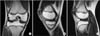
A 22-year-old male patient complained of pain and swelling on his left knee for three months with no precipitating traumatic event. From the medical history, he had undergone arthroscopic partial meniscectomy of contralateral knee at the age of 15-year-old in our hospital. He was diagnosed as having a complete medial discoid meniscus with horizontal tear and intact incomplete lateral discoid meniscus (Fig. 1, 2). The patient was treated by arthroscopic partial meniscectomy for both medial and lateral menisci. And he had no complaint about the right knee.
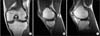
On physical examination, the patient had medial joint line tenderness and had pain on medial side in McMurray's test. A mild degree of effusion was found. Full range of motion was possible without any mechanical symptoms. No abnormalities were detected with simple radiographs. MRI of left knee showed both discoid medial and lateral menisci. Medial meniscus had horizontal tear and lateral meniscus had no tear (Fig. 3).
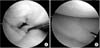
At Arthroscopy, a complete discoid medial meniscus with horizontal tear in the body and intact complete discoid lateral meniscus were found (Fig. 4). Lateral discoid meniscus didn't have hypermobility or impingement. Partial meniscectomy of medial meniscus was performed. At the follow-up one year for left knee and seven years for right knee postoperatively, the patient was asymptomatic in left knee and also in right knee. Preoperative Lysholm score of left knee was 68 but right knee had no record. At the last follow-up, Lysholm score of left knee was 97 and right knee was 94. The radiologic findings had no definite interval changes including alignment of lower limb and gaps of both medial and lateral joint space.
DISCUSSION
The incidence of discoid lateral menisci ranges from 1.5% to 15.5% and the highest incidence rate of discoid medial menisci reported is 0.3%.5) Jeannopoulos3) reported the first case of simultaneous medial and lateral discoid meniscus in the same knee in 1950. Yáñez-Acevedo6) reported one case of bilateral discoid lateral menisci and unilateral discoid medial menisci in 11-year-old girl. Kim and Lubis4) in 2010, firstly described one case of bilaterally simultaneous medial and lateral discoid menisci.
Smillie5) classified discoid menisci in three types as primitive, infantile, and intermediate. In 1979, Watanabe et al.7) classified 3 types of discoid meniscus; complete, incomplete, and Wrisberg type. The first two vary only on the degree of coverage of the meniscus. The Wrisberg type discoid menisci are hypermobile forms that lack posterior coronary ligaments and capsular attachment. In our case, with Watanabe's classification, the left knee had both medial and lateral complete discoid menisci. And the right knee, according to previous MRI and arthroscopy, had medial complete and lateral incomplete discoid menisci.
Symptoms of discoid meniscus have no specific clinical features, and the main symptoms include tenderness on the joint line, swelling, snapping, giving-way and locking.6,8) In our case, there were tenderness and swelling with effusion
Abnormal radiologic findings of discoid meniscus such as widening of joint line and cupping of the tibial plateau have been reported but our patient had no remarkable radiographic findings.4,6,9)
Therefore, diagnosis for the discoid meniscus should be confmired by either MRI or arthroscopy. Silverman et al.8) described MRI can provide accurate diagnosis and assist in pre-operative planning. A discoid meniscus is said to be present if 3 or more contiguosuasg ittal sections that are 5-mm thick show a continuity of the meniscus between the anterior and posterior horns. The presence of 2 adjacent peripheral 5-mm thick sagittal sections showing equal or nearly equal meniscal height probably indicates a discoid meniscus. Also, coronal images showing a complete meniscus, sometimes extending into the intercondylar notch, in all sections through the knee would indicate a discoid meniscus. Yilgor et al.9) reported the accuracy of MRI about discoid meniscus. The statistical analysis reveal that MRI is 100% specific and 97.8% sensitive in determining whether there is a tear in the discoid meniscus or not. MRI can predict the presence and absence of a tear with an 85.7% negative predictive value and 100% positive predictive value. In our case, all of 4-meniscus showed 3 or more contiguous sagittal sections that are 5-mm thick and both medial menisci had horizontal tear
The treatment principles of medial discoid meniscus are the same as those of lateral ones. Once validated, partial resection of torn and symptomatic discoid meniscus with arthroscopy is needed. Chen et al.10) pointed out that the surgical indication of discoid meniscus injury were similar to those of normal meniscus, and nonsurgical treatment was recommended to those without symptoms. Kim and Lubis4) reported good result of bilateral medial and lateral discoid menisci by partial meniscectomy of both torn medial menisci and preservation of intact lateral meniscus in one side. In our case, a complete discoid medial meniscus with horizontal tear in the body and intact complete discoid lateral meniscus were found. Arthroscopic partial meniscectomy of medial meniscus was performed leaving a functional residual rim of the medial meniscus. In general, the results of meniscectomy for discoid meniscus are good.4,6,10) Our patient was satisfied with the result of his surgeries and had no complaint.
 XML Download