

 PDF
PDF ePub
ePub Citation
 Print
Print


Abstract
We report on a case involving total en bloc uncinatectomy of C7 without removal of the previously inserted cage, performed on a patient with a history of previous anterior cervical discectomy and fusion without uncoforaminotomy at C5-6-7 who had persistent pain radiating to the upper extremity along with progressive weakness. Satisfactory results were achieved. This procedure should be regarded as an effective option for surgical treatment of persistent or recurrent radiculopathy caused by remaining foraminal stenosis following anterior cervical fusion, and we suggest it as a new indication for this procedure.
Figures and Tables
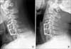
Figure 1
Lateral radiographs taken in flexion (A) and extension (B) at 14 months after the initial operation are shown. The difference in interspinous distances at C5-6 was 2.5 mm, suggesting nonunion at this level.
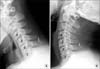
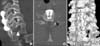
Figure 2
Computed tomography scan images are shown. (A) A left foraminal oblique image shows nonunion at C5-6 (black arrowhead) and large foraminal spurs at C6-7 (white arrowhead). (B) An axial image shows a large foraminal spur from the left uncinate process of C7 (black arrowhead).
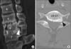
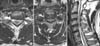
Figure 3
T2-weighted magnetic resonance images show a large foraminal spur (white arrowhead) from the left uncinate process of C7 (A), spinal cord compression between the herniated disc and slightly buckled ligamentum flavum at C4-5 (B, C), and cord signal change at C5-6 (C).
References
1. Jho HD, Jho DH. Ventral uncoforaminotomy. J Neurosurg Spine. 2007; 7:533–535.

2. Park DH, Ryu KY, Seok KS, Kang DG, Kim SC. Clinical results of microsurgical anterior foraminotomy for cervical radiculopathy. J Korean Neurosurg Soc. 2003; 34:125–129.
3. Hacker RJ, Miller CG. Failed anterior cervical foraminotomy. J Neurosurg. 2003; 98:2 Suppl. 126–130.
4. Balasubramanian C, Price R, Brydon H. Anterior cervical microforaminotomy for cervical radiculopathy--results and review. Minim Invasive Neurosurg. 2008; 51:258–262.
5. Jacobs W, Willems PC, Kruyt M, et al. Systematic review of anterior interbody fusion techniques for single- and double-level cervical degenerative disc disease. Spine (Phila Pa 1976). 2011; 36:E950–E960.
6. Shen FH, Samartzis D, Khanna N, Goldberg EJ, An HS. Comparison of clinical and radiographic outcome in instrumented anterior cervical discectomy and fusion with or without direct uncovertebral joint decompression. Spine J. 2004; 4:629–635.
7. Albert TJ, Smith MD, Bressler E, Johnson LJ. An in vivo analysis of the dimensional changes of the neuroforamen after anterior cervical diskectomy and fusion: a radiologic investigation. J Spinal Disord. 1997; 10:229–233.
8. De Palma AF, Cooke AJ. Results of anterior interbody fusion of the cervical spine. Clin Orthop Relat Res. 1968; 60:169–185.
9. Gore DR, Gardner GM, Sepic SB, Murray MP. Roentgenographic findings following anterior cervical fusion. Skeletal Radiol. 1986; 15:556–559.
10. Seo JY, Ha KY. Fate of posterior osteophytes in fused segments after anterior cervical discectomy and fusion. Spine (Phila Pa 1976). 2012; 37:741–747.
 XML Download