

 PDF
PDF ePub
ePub Citation
 Print
Print


Abstract
Monocephalus tripus tribrachius, a type of conjoined twins with one head and three upper and lower extremities, is a rare congenital disorder. To date, no long-term follow-up results of surgical procedures for this condition have been reported in Korean literature. We experienced a case of monocephalus tripus tribrachius, which had been surgically managed with an accessory lower limb disarticulation and pelvic bone reconstruction to manage this accessory limb and accompanying comorbidities in hip joint and pelvis. Subsequently, ipsilateral Syme amputation was done for intractable deformity of foot, and later, ipsilateral femoral varus derotational osteotomy was done for inadequate coverage of femoral head observed in follow-up. We report 18-year follow-up results of the procedures with a review of literatures.
Figures and Tables
Figure 1
(A) Initial plain radiograph of monocephalus tripus tribrachius. Accessory lower limb is shown on the middle portion of the pelvis and the redundant ischial bone is forming an accessory acetabular structure. (B) Schematic presentation of the initial gross appearance of the case.
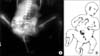
Figure 2
Plain radiograph taken at two years following accessory lower limb disarticulation and reconstruction of the pelvic bone. The wires were used to fixate both ischial bones, forming new symphysis pubis.
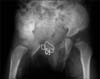
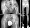
Figure 3
(A) Plain radiograph taken at 11 years of age shows good acetabular coverage and joint congruency in both hip joints. (B) The radiograph included both weight bearing whole lower limbs taken at 11 years of age. Extension-block knee brace and syme amputation prosthesis are shown. (C) The latest follow-up radiograph taken at 18 years of age.
References
1. O'Neill JA Jr, Holcomb GW 3rd, Schnaufer L, et al. Surgical experience with thirteen conjoined twins. Ann Surg. 1988; 208:299–312.
2. Spencer R. Parasitic conjoined twins: external, internal (fetuses in fetu and teratomas), and detached (acardiacs). Clin Anat. 2001; 14:428–444.

3. Mahajan JK, Devendra K, Mainak D, Rao KL. Asymmetric conjoined twins: atypical ischiopagus parasite. J Pediatr Surg. 2002; 37:E33.
4. Corona-Rivera JR, Corona-Rivera E, Franco-Topete R, Acosta-León J, Aguila-Dueñas V, Corona-Rivera A. Atypical parasitic ischiopagus conjoined twins. . J Pediatr Surg. 2003; 38:e3.
5. Votteler TP, Lipsky K. Long-term results of 10 conjoined twin separations. J Pediatr Surg. 2005; 40:618–629.
6. Lee ES, Yoo CI. A case of "monocephalus tripus dibrachius". J Korean Orthop Assoc. 1971; 6:419–424.
7. Gaine WJ, McCreath SW. Syme's amputation revisited: a review of 46 cases. J Bone Joint Surg Br. 1996; 78:461–467.
8. Sponseller PD, Jani MM, Jeffs RD, Gearhart JP. Anterior innominate osteotomy in repair of bladder exstrophy. J Bone Joint Surg Am. 2001; 83:184–193.
9. Shultz WG. Plastic repair of exstrophy of bladder combined with bilateral osteotomy of ilia. J Urol. 1958; 79:453–458.
10. Vining NC, Song KM, Grady RW. Classic bladder exstrophy: orthopaedic surgical considerations. J Am Acad Orthop Surg. 2011; 19:518–526.
 XML Download