

 PDF
PDF ePub
ePub Citation
 Print
Print


Ulnar nerve compression at the wrist is rare, but well described in the literatures.1) Compression occurs usually in Guyon's canal, a narrow passage for ulnar artery and nerve, which is made up between pisiform and hook of hamate. The possible causes are various such as ganglion, benign soft tissue masses, or anomalous muscles from hypothenar muscle. Sporadically, anatomical variants of ulnar nerve were reported as a cause of ulnar nerve symptom in the wrist.2,3,4) Herein we present a neural loop of ulnar nerve observed during ulnar nerve decompression surgery. Aberrant branch of ulnar nerve encompassing flexor carpi ulnaris (FCU) tendon is suspected to create vague ulnar side pain during active wrist flexion in the presenting case.
CASE REPORT
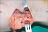
A 35-year-old male complained of vague wrist pain in ulnar side, which was especially aggravated by forceful flexion of the right wrist. Symptom started from several weeks ago without any remarkable trauma history in his wrist. He also had intermittent tingling sensation distributed to 4th and 5th finger. He demonstrated no evidence of motor deficit of the ulnar nerve. Tinel's sign was not elicited over the entire course of the ulnar nerve. Allen's test confirmed patency of the ulnar artery. X-ray films of the wrist were normal. After a few weeks of conservative treatment, we performed electromyogram and nerve conduction velocity test since there was no improvement of symptom. In the test there is no definite neuroelectrophysiological abnormality indicating peripheral neuropathy or radiculopathy. Inching test was not performed to find exact location of the entrapment since the result of conventional electrophysiological study was normal. Ultrasonography neither revealed any specific local abnormality in wrist and elbow. Due to minimal response on further duration of conservative treatment, the exploration of Guyon's canal was carried out with the impression of distal ulnar nerve compression at the wrist. The purpose of surgery was exploration and release of Guyon's canal. The exploration revealed a neural loop of ulnar nerve relatively proximal to pisiform, which encompassed FCU tendon (Fig. 1). This aberrant fascicle ran proximally encompassing FCU and rejoined to major trunk of ulnar nerve at approximately 2 cm proximal from the start point. We assumed that a neural loop could be one of sources eliciting a vague ulnar side pain during active wrist flexion in this case. Release of Guyon's canal, epineurolysis of aberrant neural loop, and release of FCU from surrounding tissues were performed (Fig. 2). Tight association between FCU and neural loop was relieved after adhesiolysis and neurolysis, resulting in redundant neural loop around FCU. The transection and repair of FCU to correct encompassing by neural loop was not attempted. At two years follow-up, he is completely free of his ulnar side wrist pain and intermittent tingling sensation.
DISCUSSION
Neural loops of ulnar nerve were rarely reported in English literatures. This rare anatomic variant could be arbitrarily divided into two distinct groups by its location, as proximal or distal from pisiform.4,5) Since compression neuropathy usually occurs distal to the pisiform, some anatomical studies focused on neural loop distal to pisiform. Bergfield and Aulicino2) reported that they encountered distal neural loop on three occasions during neurolysis of the ulnar nerve through Guyon's canal in clinical situation. This neural loop surrounds the hook of hamate consistently and rejoins the nerve distally deep in the palm (Fig. 3A, 3B). Subsequently Rogers et al.5) reported in their cadaveric study that they found this kind of neural loop on 7 cases out of 77 cadaver wrist dissections (9%), all the location was at deep motor branch.
Neural loop proximal to the pisiform has been rarely reported. Dodds et al.6) reported a variant in which additional branch from dorsal cutaneous branch of ulnar nerve was re-joined to superficial branch of ulnar nerve from cadaveric study (1 case out of 58 paired cadaver wrists [1.7%], Fig. 3C). Approximately 8 cm proximal to the pisiform the ulnar nerve gave off a branch, which passed deep to FCU, and joining the superficial branch of the ulnar nerve at the distal edge of the pisiform. Bonnel and Vila7) reported a similar case in cadaveric study (1 case in 50 hands). Neural loop was formed between dorsal cutaneous branch and ulnar proper palmar digital nerve of the little finger.
Clinically relevant reports about proximal neural loop are also scant. There were two case reports of the symptomatic neural loop, which had been penetrating FCU, not encompassing (Fig. 3D). In one case, aberrant FCU insertion was concomitantly observed with neural loop penetrating FCU.8) After relocation of aberrant FCU insertion and epineurolysis, the symptom of the patient was completely relieved. Kang et al.9) also reported the similar neural loop penetrating FCU and subsequent symptom relief after similar operation. In 2005, Musthyala and Jones4) reported the most similar form of neural loop with ours. Thirty-seven year-old female who suffered from ulnar side-wrist pain was explored and neural loop encompassing FCU was found (Fig. 3E). In contrast to our case, they transected FCU tendon at musculocutaneous junction to release neural loop from FCU tendon. After the neural loop was freed, the tendon was re-united, and subsequently symptoms improved with time. In our case, simple decompression of neural loop and Guyon canal alone was sufficient to produce symptom relief for 2 years.
However, there is still lack of direct cause and effect relationship between the neural loop and the symptom of patient. Since we conducted the combination surgery including 1) Guyon's canal release, 2) epineurolysis of ulnar nerve, and 3) release of FCU, it is unclear which procedure was corresponding for symptom relief. Because the conservative management was failed in prolonged period, we performed exploratory surgery considering Guyon's canal release as a main plan, we incidentally encountered this anomaly. Considering the dynamic nature of symptom in this patient, we further conducted epineurolysis of ulnar nerve and release of FCU to eliminate any possibility of ulnar nerve compression, either static or dynamic. We speculated that the ulnar nerve compression or irritation by forceful wrist flexion might be cause of the ulnar side wrist pain as the patients with radial tunnel syndrome experience vague forearm pain along the course of posterior interosseous nerve.
With regard to the ulnar side wrist pain, which is suspicious for ulnar compression syndrome at the wrist level, the surgeon should always suspect anomalous nerve branch as source of compressive neuropathic pain. Despite of almost normal neuroelectrophysiological study, aberrant ulnar nerve variants might elicit symptoms in relation with active motion of nearby flexor tendon.


 XML Download