

 PDF
PDF ePub
ePub Citation
 Print
Print


Weight lifting usually involves lifting a weight heavier than one's own body above the head level by using fast and strong elbow joint extensibility with a pronated forearm and a wide, weight-bearing grip, and this may lead to strong valgus force on the elbow joint. Chronic repetitive valgus stress during exercise and abrupt resisted extension force on lifting could cause injuries at the medial side of the elbow joint. We experienced a case of chronic complete medial collateral ligament and common flexor tendon rupture in a 16-year-old female weight lifting athlete and she was treated with surgical reconstruction and she was evaluated with serial ultrasonographic examinations. We report here on the usefulness of an ultrasonographic examination for identifying the integrity of the reconstructed ligament and supporting the rehabilitation program.
CASE REPORT
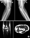
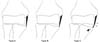
A 16-year-old high school female weight-lifter suffered a valgus overload injury with a popping sound during a weight lifting competition 6 months prior to visiting our outpatient clinic. She did not received specific treatment other than a few days rest regardless of the slight swelling and bruise on her left elbow joint area. She noticed progressive weakness of her elbow flexion power and she couldn't elevate a barbell after experiencing a similar type of recurrent injury during exercise 1 month ago. She was an elite weight lifter with a 3 years career and she averaged 70 kg for the snatch event and 90 kg for the clean and jerk event. On physical examination, there were mild tenderness on the medial elbow joint and increased valgus laxity compared to the contralateral side. The medial joint width on the valgus stress view was 7.0 mm on the left elbow and 4.9 mm on the right side. Magnetic resonamce imaging (MRI) examination revealed a torn medial collateral ligament and a retracted common flexor tendon (Fig. 1). The initial ultrasonographic findings revealed a torn medial collateral ligament and common flexor tendon with hypoechogenic areas of discontinuity. The angular deformity on the Sasaki classification1) was type C and the gap of the elbow joint (horizontal distance) was 8.4 mm and the lateral movement of the elbow joint (vertical distance) was -1.2 mm (Fig. 2, 3).
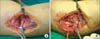
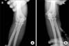
She underwent ulnar collateral ligament reconstruction using the palmaris longus tendon and common flexor tendon repair (Fig. 4). After 6 weeks' immobilization with a long arm cast, passive range of motion exercise and isometric exercise were begun. Active range of motion exercise and isotonic exercise were started after eight weeks within the range of the pain free motion arc and weight. An isokinetic exercise training program was started 3 months after the operation and weight lifting exercise permitted after 6 months. On the contrary to our rehabilitation program, she started lifting exercise 4 months after the operation to attend local competition. She complained of mild pain and tenderness on the medial joint space at 6 months' follow-up. Based on 6 months' ultrasonographic findings, we prohibited vigorous exercise program for another 1 month and restarted strengthening exercise seven months after operation. The patient recovered 64% and 78% of the preinjury level records (45 kg for the snatch event and 70 kg for the clean and jerk event) at 9 months after operation and more than 90% of her pre-injury strength (65 kg for the snatch event and 90 kg for the clean and jerk event) at 1 year after operation. Mayo elbow performance score was 60 in initial and improved to 85 at 3 months and 6 months after operation, and 92 at one year follow-up examination. There were no limitation of elbow motion compared to nonoperated side and the medial joint width on the valgus stress view revealed an intact medial joint space compared to normal side (Fig. 5).
1. Ultrasonographic findings
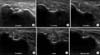
The serial ultrasonographic evaluation was done at the initial examination as well as at 6 weeks, 3 months, 6 months, 9 months, and 1 year after the operation to assess and evaluate the condition of the repaired ligament as well as its functional recovery. For the ultrasonographic examination, the patient was positioned in the supine position, with the arm undergoing examination stretched across the bed. After the arm was 90o in external rotation, the elbow joint bent at 70o, and the forearm in a neutral position, gravity bearing stress was put on the forearm in order to cause tension in the medial portion of the elbow joint. The horizontal and vertical distances were 8.4 mm, 2.0 mm, 2.6 mm, 2.8 mm, 2.7 mm, 2.7 mm, and -1.2 mm, 2.4 mm, 1.0 mm, 0.0 mm, 0.0 mm, 0.0 mm, respectively (Table 1). The angular deformity was type A at 6 weeks and 3 months after the operation, and type B at 6 months, 9 months and 1 year after the operation. The ultrasonographic findings of the 6 weeks after operation revealed that the reconstructed ligament was maintained with a reduced distance of the vertical and horizontal spaces. The fibrillar pattern of the ligament was smooth and parallelized at 3 months follow-up, but increased inhomogeneity and swelling around the reconstructed ligament and a type B nature joint space were identified at the 6 months evaluation, which might have been influenced by the increased activity and weight training. We restricted weight lifting exercise for 1 month and restarted lifting exercise 7 months after operation. A clearer hyperechogenic fibrillar pattern of the reconstructed ligament could be identified at the 9 months and 1 year follow-up in spite of the sustained hypoechogenecity around the reconstructed ligament and the hyperplasia of the surrounding soft tissue (Fig. 6).
DISCUSSION
Weight lifting needed strong muscular power for lifting as well as valgus stability for maintaining balance and control of the barbell. During the concentric movement of the bench press, there is an initial high-power push after chest contact, immediately followed by a characteristic area of low power, the so-called 'sticking region'. The decline in power during the initial acceleration phase appears to be a factor in a failed lift attempt at the sticking point during high intensity lifting or fatigue repeat repetition training, and this increases the risk of injuries.2) Medial joint injuries can be identified with careful history taking and a thorough physical examination and they can be confirmed with radiologic imaging studies, including simple and stress radiography, arthrography, computerized tomography, MRI and dynamic ultrasonographic examination.1,3,4,5,6,7,8) MRI can be a useful diagnostic study for medial collateral ligament injury and injury to the surrounding soft tissue structures in both the acute and chronic conditions, but its usefulness as a screening test for athletes might be limited due to MRI's high cost. According to Timmerman et al.,8) MRI showed 57% sensitivity and 100% specificity for diagnosing medial collateral ligament injury. However Thompson et al.9) reported 79% positive results and 21% false negative results for patients who received reconstructive surgery. The effectiveness of dynamic ultrasonography to confirm ulnar collateral ligament injury was recently reported by several authors.1,3,7) Sasaki et al.1) reported on performing dynamic ultrasonography for evaluating medial joint laxity and medial collateral ligament injury and classified three types of angular changes of the medial collateral ligament and measurement of the medial joint space. They revealed that the medial elbow joint gap and angular deformity were more increased in the dominant arm of 30 males baseball pitchers. But there have been no reports on the usefulness of ultrasonographic study for identifying ligament injury and serial examination for evaluating the integrity of the reconstructed ligament to support a rehabilitation program.
Actually, presented patient started weight lifting exercise before 6 months after the operation to attend local competition, because general symptoms get improved and she had pressures to return to the pre-injury level of exercise. Serial ultrasonographic examination could be used as an impressive imaging study identifying the condition of the reconstructed ligament to the athletes regardless of improved pain or strength after operation. Based on 6 month's ultrasonographic findings, we delayed vigorous exercise program for 1 month. The patient recovered 64% and 78% of the preinjury level records (45 kg for the snatch event and 70 kg for the clean and jerk event) at 9 months after operation and more than 90% of her pre-injury strength (65 kg for the snatch event and 90 kg for the clean and jerk event) at 1 year after operation. Ultrasonographic examination at 1 year revealed that the integrity and thickness of the reconstructed ligament were preserved with the type B angle of the elbow joint. But, there were hypoechogenesity around the reconstructed ligament and hyperplasia of the surrounding soft tissue, which required that the patient be aware of the risk of reinjury and that she should continue her rehabilitation exercise. Young athletes of growing age needed regular examination during training programs to identify injuries and so administer early treatment. An ultrasonographic examination could be a useful diagnostic and follow-up study for evaluating musculoskeletal injuries of athletes because it is a noninvasive, well tolerated and quick study with immediate results.


 XML Download