

 PDF
PDF ePub
ePub Citation
 Print
Print


Abstract
Angiolymphoid hyperplasia with eosinophilia (ALHE) and Kimura's disease were classified as same disease in the past. Since there are many different clinical and histopathological characteristics that warranted their distinction, they are classified as different disease now. Six cases of Kimura's disease in upper extremity have been reported in Korean literature but ALHE in upper extremity has not been reported yet. We experienced a case of surgically treated ALHE in the upper arm and report this case with review of literature.
Figures and Tables
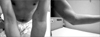
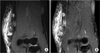
Figure 2
Magnetic resonance images show 2 enlarged lymph nodes on medial aspect of left upper arm, high signal intensity on fat-suppressed T2-weighted image (A) and marginal enhancement on Gadolinium enhanced fat-suppressed T1-weighted image (B). Severe edema and inflammatory change in surrounding subcutaneous tissue are also observed.
References
1. Ramchandani PL, Sabesan T, Hussein K. Angiolymphoid hyperplasia with eosinophilia masquerading as Kimura disease. Br J Oral Maxillofac Surg. 2005. 43:249–252.

2. Chong WS, Thomas A, Goh CL. Kimura's disease and angiolymphoid hyperplasia with eosinophilia: two disease entities in the same patient: case report and review of the literature. Int J Dermatol. 2006. 45:139–145.
3. Kim HT, Szeto C. Eosinophilic hyperplastic lymphogranuloma comparison with Mikulicz's disease. Chin Med J. 1937. 23:699–700.
4. Kimura T, Yoshimura S, Ishikawa E. Abnormal granuloma with proliferation of lymphoid tissue. Trans Soc Pathol Jpn. 1948. 37:179–180.
5. Wells GC, Whimster IW. Subcutaneous angiolymphoid hyperplasia with eosinophilia. Br J Dermatol. 1969. 81:1–14.
6. Rosai J, Gold J, Landy R. The histiocytoid hemangiomas. A unifying concept embracing several previously described entities of skin, soft tissue, large vessels, bone, and heart. Hum Pathol. 1979. 10:707–730.
7. Kuo TT, Shih LY, Chan HL. Kimura's disease. Involvement of regional lymph nodes and distinction from angiolymphoid hyperplasia with eosinophilia. Am J Surg Pathol. 1988. 12:843–854.
8. Googe PB, Harris NL, Mihm MC Jr. Kimura's disease and angiolymphoid hyperplasia with eosinophilia: two distinct histopathological entities. . J Cutan Pathol. 1987. 14:263–271.
9. Lenk N, Artüz F, Kulaçoğlu S, Alli N. Kimura's disease. Int J Dermatol. 1997. 36:437–439.
10. Hui PK, Chan YW. Kimura's disease--treatment with steroid. Histopathology. 1990. 17:286–287.
 XML Download