

 PDF
PDF ePub
ePub Citation
 Print
Print


Abstract
Purpose
The aim of this study was to evaluate the correlation between the 3-dimensional (D) notch volume and the 2D notch width and notch shape as measured on magnetic resonance imaging (MRI), between subjects with anterior cruciate ligament (ACL) injury and those without ACL injury.
Materials and Methods
Knee MRI images were taken from 72 subjects with ACL injury and 80 subjects without ACL injury (January 2007 to January 2012; Gyeongsang National University Hospital, Jinju, Korea). We measured 3D notch volume and 2D notch width and notch shape. The measured values from MRI figures between ACL-injured subjects and non-ACL-injured subjects were compared and analyzed. These measurements (notch width, notch ratio) were correlated to notch volume. Both intra-observer reliability and inter-observer reliability were calculated.
Figures and Tables
Figure 1
T2-weighted axial images, The borders of the notch are outlined. The posterior border of the notch: a line between the two points on the inside of the femoral condyles where the cartilage ends. (A) First image with both femoral condyles visible, as indicated by the articular cartilage. (B) Last image with continuity between the medial and lateral femoral condyles. The notch volume is calculated by summation of the area of all images within the defined femoral notch and multiplication by the slice thickness (3 mm).
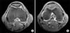
Figure 2
T1-weighted coronal image. A line is shown along the most inferior part of the femoral condyles and a line parallel to this line at an intermediate image among those obtained where the popliteal grooves can be seen, where the notch width was measured.
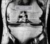
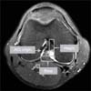
Figure 3
T2-weighted axial image, Measurement of the length of the base and height of the intercondylar notch at the anterior cruciate ligament (ACL) origin, ratio (height/base) was performed.

References
1. Cha JH, Lee SH, Shin MJ, Choi BK, Bin SI. Relationship between mucoid hypertrophy of the anterior cruciate ligament (ACL) and morphologic change of the intercondylar notch: MRI and arthroscopy correlation. Skeletal Radiol. 2008; 37:821–826.

2. Gwinn DE, Wilckens JH, McDevitt ER, Ross G, Kao TC. The relative incidence of anterior cruciate ligament injury in men and women at the United States Naval Academy. Am J Sports Med. 2000; 28:98–102.
3. Oliphant JG, Drawbert JP. Gender differences in anterior cruciate ligament injury rates in wisconsin intercollegiate basketball. J Athl Train. 1996; 31:245–247.
4. Shelbourne KD, Davis TJ, Klootwyk TE. The relationship between intercondylar notch width of the femur and the incidence of anterior cruciate ligament tears. A prospective study. Am J Sports Med. 1998; 26:402–408.
5. Shelbourne KD, Facibene WA, Hunt JJ. Radiographic and intraoperative intercondylar notch width measurements in men and women with unilateral and bilateral anterior cruciate ligament tears. Knee Surg Sports Traumatol Arthrosc. 1997; 5:229–233.
6. Stijak L, Radonjić V, Aksić M, Filipović B, Sladojević M, Santrac-Stijak G. Correlation between femur's length and morphometric parameters of distal femur important in rupture anterior cruciate ligament. Acta Chir Iugosl. 2009; 56:61–66.
7. Uhorchak JM, Scoville CR, Williams GN, Arciero RA, St Pierre P, Taylor DC. Risk factors associated with noncontact injury of the anterior cruciate ligament: a prospective four-year evaluation of 859 West Point cadets. Am J Sports Med. 2003; 31:831–842.
8. Dienst M, Schneider G, Altmeyer K, et al. Correlation of intercondylar notch cross sections to the ACL size: a high resolution MR tomographic in vivo analysis. Arch Orthop Trauma Surg. 2007; 127:253–260.
9. Hernigou P, Garabedian JM. Intercondylar notch width and the risk for anterior cruciate ligament rupture in the osteoarthritic knee: evaluation by plain radiography and CT scan. Knee. 2002; 9:313–316.
10. Murshed KA, Ciçekcibaşi AE, Karabacakoğlu A, Seker M, Ziylan T. Distal femur morphometry: a gender and bilateral comparative study using magnetic resonance imaging. Surg Radiol Anat. 2005; 27:108–112.
11. Tillman MD, Smith KR, Bauer JA, Cauraugh JH, Falsetti AB, Pattishall JL. Differences in three intercondylar notch geometry indices between males and females: a cadaver study. Knee. 2002; 9:41–46.
12. Ireland ML, Ballantyne BT, Little K, McClay IS. A radiographic analysis of the relationship between the size and shape of the intercondylar notch and anterior cruciate ligament injury. Knee Surg Sports Traumatol Arthrosc. 2001; 9:200–205.
13. Charlton WP, St John TA, Ciccotti MG, Harrison N, Schweitzer M. Differences in femoral notch anatomy between men and women: a magnetic resonance imaging study. Am J Sports Med. 2002; 30:329–333.
14. Lombardo S, Sethi PM, Starkey C. Intercondylar notch stenosis is not a risk factor for anterior cruciate ligament tears in professional male basketball players: an 11-year prospective study. Am J Sports Med. 2005; 33:29–34.
15. Herzog RJ, Silliman JF, Hutton K, Rodkey WG, Steadman JR. Measurements of the intercondylar notch by plain film radiography and magnetic resonance imaging. Am J Sports Med. 1994; 22:204–210.
16. van Eck CF, Kopf S, van Dijk CN, Fu FH, Tashman S. Comparison of 3-dimensional notch volume between subjects with and subjects without anterior cruciate ligament rupture. Arthroscopy. 2011; 27:1235–1241.
17. Vrooijink SH, Wolters F, Van Eck CF, Fu FH. Measurements of knee morphometrics using MRI and arthroscopy: a comparative study between ACL-injured and non-injured subjects. Knee Surg Sports Traumatol Arthrosc. 2011; 19:Suppl 1. S12–S16.
18. Tashman S, Kopf S, Fu FH. The kinematic basis of ACL reconstruction. Oper Tech Sports Med. 2008; 16:116–118.
19. Shen W, Forsythe B, Ingham SM, Honkamp NJ, Fu FH. Application of the anatomic double-bundle reconstruction concept to revision and augmentation anterior cruciate ligament surgeries. J Bone Joint Surg Am. 2008; 90:Suppl 4. 20–34.
20. Domzalski M, Grzelak P, Gabos P. Risk factors for anterior cruciate ligament injury in skeletally immature patients: analysis of intercondylar notch width using magnetic resonance imaging. Int Orthop. 2010; 34:703–707.
21. Van Eck CF, Martins CA, Kopf S, Lertwanich P, Fu FH, Tashman S. Correlation between the 2-dimensional notch width and the 3-dimensional notch volume: a cadaveric study. Arthroscopy. 2011; 27:207–212.
22. van Eck CF, Martins CA, Vyas SM, Celentano U, van Dijk CN, Fu FH. Femoral intercondylar notch shape and dimensions in ACL-injured patients. Knee Surg Sports Traumatol Arthrosc. 2010; 18:1257–1262.
 XML Download