

 PDF
PDF ePub
ePub Citation
 Print
Print


Abstract
In elderly patients, the first incidence of gout often affects the distal interphalangeal joint (DIP) and usually subsides without specific treatment after about 7 days. A 61-year-old male was presented to our clinic with a 10-day history of tenderness and swelling in his index DIP, which was initially diagnosed as cellulitis. After a skin incision was made to drain the lesion, typical tophaceous deposits were observed around the extensor apparatus, flexor tendons, and joint capsule. The tophi were meticulously removed in order to minimize the injury to its surrounding structures, after which the joint fluid was aspirated. There was no history of gout, laboratory findings suggesting tophaceous gout, or apparent predisposing factors in the patient's history. Pathology confirmed tophaceous deposits and negative birefringent crystals, and the patient has been managed on allopurinol for post-operative six months.
Figures and Tables
Figure 1
A simple radiograph of acute attack of gout in the distal interphalangeal joint at left index finger. There were no specific findings, except marked soft tissue swelling.
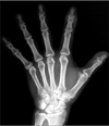
Figure 2
(A) Appearance of the patient's dorsal left hand at presentation. (B) Surgical site after lateral skin incision with the typical appearance of a gouty tophus. The tophus was easily separated from the extensor apparatus, flexor tendon sheath, and capsule. Note the thin, transparent appearance of the capsule above the extensor apparatus. (C) Gross finding of tophaceous deposits.
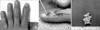
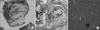
Figure 3
(A) Photomicrograph of a specimen demonstrating gouty tophus with synovial tissue (H&E stain, ×40). (B) Amorphous central material deposits (asterisks) surrounded by inflammatory cells (arrows) (H&E stain, ×200). (C) Needle-shaped urate crystal from distal interphalangeal joint fluid showing negative birefringence under compensated polarized light microscopy.

References
1. Wallace SL, Singer JZ. Therapy in gout. Rheum Dis Clin North Am. 1988. 14:441–457.

2. Hernández-Cortés P, Caba M, Gómez-Sánchez R, Gómez-Morales M. Digital flexion contracture and severe carpal tunnel syndrome due to tophaceus infiltration of wrist flexor tendon: first manifestation of gout. Orthopedics. 2011. 34:e797–e799.
3. Mittag F, Wuenschel M. Giant gouty tophi of the hand and wrist. Orthopedics. 2011. 34:e790–e792.
4. Thissen CA, Frank J, Lucker GP. Tophi as first clinical sign of gout. Int J Dermatol. 2008. 47:Suppl 1. 49–51.
5. Urano W, Yamanaka H, Tsutani H, et al. The inflammatory process in the mechanism of decreased serum uric acid concentrations during acute gouty arthritis. J Rheumatol. 2002. 29:1950–1953.
6. Corrado A, D'Onofrio F, Santoro N, Melillo N, Cantatore FP. Pathogenesis, clinical findings and management of acute and chronic gout. Minerva Med. 2006. 97:495–509.
7. Dhoble A, Balakrishnan V, Smith R. Chronic tophaceous gout presenting as acute arthritis during an acute illness: a case report. Cases J. 2008. 1:238.
 XML Download