

 PDF
PDF ePub
ePub Citation
 Print
Print


Abstract
Purpose
Third generation shoulder arthroplasty is widely performed nowadays; however, few studies on the anatomy of the proximal humerus in the Korean population have been reported. The authors have attempted to review the anatomy of the proximal humerus.
Materials and Methods
The study sample consisted of 100 humeri of patients with a mean age of 48 years (range of 17 to 83 years) who underwent computed tomography imaging between January 2009 and October 2011 at Myongji Hospital. Diameter of the articular surface, head thickness, radius of curvature, head inclination, head to tuberosity height, bicipital groove-shaft angle, lateral angle, medial offset and posterior offset were analyzed. Results were compared depending on age and gender.
Results
Mean values of diameter of the articular surface was 42.70±3.57 mm, head thickness was 14.3±2.0 mm, and radius of curvature was 22.50±1.97 mm; these three variables showed significant sex differences. Head inclination was measured as 130.00±4.28 degrees, head to tuberosity height was 7.50±0.99 mm, bicipital groove-shaft angle was 6.60±0.92 degrees, and lateral angle was 163.40±4.05 degrees. Mean medial and posterior offset were 5.2±2.1 mm and 3.1±1.8 mm, respectively.
Figures and Tables
Figure 1
(A) The diameter of the articular surface (a) measured a distance between cartilage edges of the humeral head in the coronal plane. The distance from the peak of the articular surface to the diameter of the articular surface (a) is head thickness (b). The radius of curvature (c) measured the radius of the humeral head in the sagittal plane. (B) Angle (d) formed between the perpendicular line to the diameter of the articular surface and axis of the humeral shaft in the coronal plane.
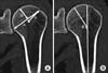
Figure 2
(A) The distance (e) from the peak of the humeral head to the peak of greater tuberosity in the coronal plane. (B) Angle (f) formed between the axis of the humeral shaft to the bicipital groove in the 3 dimensional reconstruction image. (C) Angle (g) formed between lateral contours of the proximal humerus. (D) Humeral head offset distance. The metaphyseal cylinder center and the humeral head center is shown. The distance between the metaphyseal cylinder center and the humeral head center is characterized by medial offset (h), posterior offset (i).
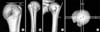


Table 6
Morphometric Data of Korean Humerus Compared with Caucasian Data

*Women≥65 years. †Reproduced from Boileau and Walch. J Bone Joint Surg Br. 1997;79:857-65 with permission of the copyright holder.2)
References
1. Pearl ML, Kurutz S. Geometric analysis of commonly used prosthetic systems for proximal humeral replacement. J Bone Joint Surg Am. 1999; 81:660–671.

2. Boileau P, Walch G. The three-dimensional geometry of the proximal humerus. Implications for surgical technique and prosthetic design. J Bone Joint Surg Br. 1997; 79:857–865.
3. Chun JM, Chung ER, Kim KY. Measurement of proximal humerus in Korean adult skeleton. J Korean Orthop Assoc. 1999; 34:219–226.
4. Jahng JS, Wee KM, Lee KH. The morphological study on the proximal part of the humerus in the Korean adults. J Korean Orthop Assoc. 1983; 18:507–512.
5. Court-Brown CM, Garg A, McQueen MM. The epidemiology of proximal humeral fractures. Acta Orthop Scand. 2001; 72:365–371.
6. Landis JR, Koch GG. The measurement of observer agreement for categorical data. Biometrics. 1977; 33:159–174.
7. Oh JH, Song BW. The current state of total shoulder arthroplasty. Clin Should Elbow. 2011; 14:253–261.
8. Bohsali KI, Wirth MA, Rockwood CA Jr. Complications of total shoulder arthroplasty. J Bone Joint Surg Am. 2006; 88:2279–2292.
9. Nuttall D, Haines JF, Trail IA. The effect of the offset humeral head on the micromovement of pegged glenoid components: a comparative study using radiostereometric analysis. J Bone Joint Surg Br. 2009; 91:757–761.
10. Pearl ML, Volk AG. Retroversion of the proximal humerus in relationship to prosthetic replacement arthroplasty. J Shoulder Elbow Surg. 1995; 4:286–289.
11. Edelson G. Variations in the retroversion of the humeral head. J Shoulder Elbow Surg. 1999; 8:142–145.
 XML Download