

 PDF
PDF ePub
ePub Citation
 Print
Print


Abstract
Pseudoaneurysm resulting from vascular impingement by an osteochondroma is extremely rare. The authors report on the case of a 16-year-old male who had a brachial artery pseudoaneurysm and vessel rupture associated with a humeral osteochondroma. This case suggests that pseudoaneurysm should be considered for the differential diagnosis in patients with soft tissue masses and a cuspidal osteochondroma located near the neurovascular bundle and recommends Doppler sonography or angiography.
Figures and Tables
Figure 1
(A) Plain radiograph shows a left proximal humeral osteochondroma. (B) Three-dimentional reconstructed computed tomography (CT) image shows a medially protruded humeral osteochondroma. (C) Axial CT image demonstrates that this osteochondroma has a sharp beak toward the brachial neurovascular bundle.
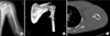
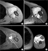
Figure 2
(A) The shoulder T1 and (B) T2-weighted magnetic resonance (MR) axial images demonstrate soft tissue mass. (C) Enhance T1-weighted MR axial image shows pseudoaneurysm (arrow) arising from the compressed brachial artery which demonstrate a strong enhancement. (D) T2-weighted MR shows that this soft tissue mass has high signal intensity along the intermuscular septum.
References
1. Eschelman DJ, Gardiner GA Jr, Deely DM. Osteochondroma: an unusual cause of vascular disease in young adults. J Vasc Interv Radiol. 1995; 6:605–613.

2. Scotti C, Marone EM, Brasca LE, et al. Pseudoaneurysm overlying an osteochondroma: a noteworthy complication. J Orthop Traumatol. 2010; 11:251–255.
3. Vasseur MA, Fabre O. Vascular complications of osteochondromas. J Vasc Surg. 2000; 31:532–538.
4. Kim YJ, Baek WK, Kim JY, et al. Pseudoaneurysm of the popliteal artery mimicking tumorous condition. J Korean Surg Soc. 2011; 80:Suppl 1. S71–S74.
5. Bilotta W, Walker H, McDonald DJ, Sundaram M. Case report 651: thrombosed, leaking popliteal aneurysm. Skeletal Radiol. 1991; 20:71–72.
6. Mann HA, Hilton A, Goddard NJ, Smith MA, Holloway B, Lee CA. Synovial sarcoma mimicking haemophilic pseudotumour. Sarcoma. 2006; 2006:27212.
7. Hermann G, Abdelwahab IF, Miller TT, Klein MJ, Lewis MM. Tumour and tumour-like conditions of the soft tissue: magnetic resonance imaging features differentiating benign from malignant masses. Br J Radiol. 1992; 65:14–20.
8. Tobias AM, Chang B. A rare brachial artery pseudoaneurysm 13 years after excision of a humeral osteochondroma. Ann Plast Surg. 2004; 52:419–422.
9. Gerrand CH. False aneurysm and brachial plexus palsy complicating a proximal humeral exostosis. J Hand Surg Br. 1997; 22:413–415.
10. Longo JM, Rodríguez-Cabello J, Bilbao JI, Aquerreta JD, Ruza M, Mansilla F. Popliteal vein thrombosis and popliteal artery compression complicating fibular osteochondroma: ultrasound diagnosis. J Clin Ultrasound. 1990; 18:507–509.
 XML Download