

 PDF
PDF ePub
ePub Citation
 Print
Print


Abstract
Purpose
The purpose of this study was to evaluate cross-sectional area of the median nerve using ultrasound in patients with carpal tunnel syndrome before and after endoscopic intervention, and to verify the level at which it can be used in prediction of outcome.
Materials and Methods
A prospective study was conducted in 21 patients who underwent endoscopic carpal tunnel release from March 2011 to March 2012. Median nerve cross-sectional area was measured before the operation and three months after the operation at the level of lunate, pisiform and hamate. The Boston questionnaire was evaluated before the operation and three months after the operation, and then allocated as two groups (group I: symptom improvement of more than 25%, group II: symptom improvement less than 25%). Then, differences of cross-sectional area between preoperative measurement and postoperative measurement on three levels were compared between the two groups.
Figures and Tables
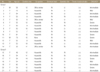
 | Figure 1Ultrasonographic measurement of median nerve (white arrow, circle) cross-sectional area at three different levels. (A) Lunate (L) level, (B) pisiform (P) level, (C) hamate (H) level.
|
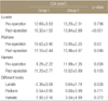
Table 1
Patient Data
*Based on classification of Stevens.12) EMG, electromyography; NCV, nerve conduction velocity; M, male; F, female; Rt, right; Lt, left.
![]()

References
1. Buchberger W, Schön G, Strasser K, Jungwirth W. High-resolution ultrasonography of the carpal tunnel. J Ultrasound Med. 1991; 10:531–537.

2. Phalen GS. The carpal-tunnel syndrome. Seventeen years' experience in diagnosis and treatment of six hundred fifty-four hands. J Bone Joint Surg Am. 1966; 48:211–228.
3. Chen P, Maklad N, Redwine M, Zelitt D. Dynamic high-resolution sonography of the carpal tunnel. AJR Am J Roentgenol. 1997; 168:533–537.
4. Buchberger W, Judmaier W, Birbamer G, Lener M, Schmidauer C. Carpal tunnel syndrome: diagnosis with high-resolution sonography. AJR Am J Roentgenol. 1992; 159:793–798.
5. El-Karabaty H, Hetzel A, Galla TJ, Horch RE, Lücking CH, Glocker FX. The effect of carpal tunnel release on median nerve flattening and nerve conduction. Electromyogr Clin Neurophysiol. 2005; 45:223–227.
6. Lee CH, Kim TK, Yoon ES, Dhong ES. Postoperative morphologic analysis of carpal tunnel syndrome using high-resolution ultrasonography. Ann Plast Surg. 2005; 54:143–146.
7. Hammer HB, Hovden IA, Haavardsholm EA, Kvien TK. Ultrasonography shows increased cross-sectional area of the median nerve in patients with arthritis and carpal tunnel syndrome. Rheumatology (Oxford). 2006; 45:584–588.
8. Colak A, Kutlay M, Pekkafali Z, et al. Use of sonography in carpal tunnel syndrome surgery. A prospective study. Neurol Med Chir (Tokyo). 2007; 47:109–115.
9. Abicalaf CA, de Barros N, Sernik RA, et al. Ultrasound evaluation of patients with carpal tunnel syndrome before and after endoscopic release of the transverse carpal ligament. Clin Radiol. 2007; 62:891–894.
10. Katz JN, Losina E, Amick BC 3rd, Fossel AH, Bessette L, Keller RB. Predictors of outcomes of carpal tunnel release. Arthritis Rheum. 2001; 44:1184–1193.
11. Agee JM, McCarroll HR Jr, Tortosa RD, Berry DA, Szabo RM, Peimer CA. Endoscopic release of the carpal tunnel: a randomized prospective multicenter study. J Hand Surg Am. 1992; 17:987–995.
12. Stevens JC. AAEE minimonograph #26: the electrodiagnosis of carpal tunnel syndrome. Muscle Nerve. 1987; 10:99–113.
13. Levine DW, Simmons BP, Koris MJ, et al. A self-administered questionnaire for the assessment of severity of symptoms and functional status in carpal tunnel syndrome. J Bone Joint Surg Am. 1993; 75:1585–1592.
14. Naranjo A, Ojeda S, Araña V, et al. Usefulness of clinical findings, nerve conduction studies and ultrasonography to predict response to surgical release in idiopathic carpal tunnel syndrome. Clin Exp Rheumatol. 2009; 27:786–793.
15. Akelman E, Weiss AP. Carpal tunnel syndrome. Etiology and endoscopic treatment. Orthop Clin North Am. 1995; 26:769–778.
16. Omer GE Jr. Median nerve compression at the wrist. Hand Clin. 1992; 8:317–324.
17. Fornage BD, Rifkin MD. Ultrasound examination of the hand and foot. Radiol Clin North Am. 1988; 26:109–129.
18. Fornage BD. Peripheral nerves of the extremities: imaging with US. Radiology. 1988; 167:179–182.
19. Middleton WD, Kneeland JB, Kellman GM, et al. MR imaging of the carpal tunnel: normal anatomy and preliminary findings in the carpal tunnel syndrome. AJR Am J Roentgenol. 1987; 148:307–316.
20. Mesgarzadeh M, Schneck CD, Bonakdarpour A, Mitra A, Conaway D. Carpal tunnel: MR imaging. Part II. Carpal tunnel syndrome. Radiology. 1989; 171:749–754.
21. Lin YM, Lee TS. Cutoff point of 0.10 cm2 appropriate for both hands. Radiology. 2005; 234:642.
22. Lee D, van Holsbeeck MT, Janevski PK, Ganos DL, Ditmars DM, Darian VB. Diagnosis of carpal tunnel syndrome. Ultrasound versus electromyography. Radiol Clin North Am. 1999; 37:859–872.
23. Mondelli M, Filippou G, Aretini A, Frediani B, Reale F. Ultrasonography before and after surgery in carpal tunnel syndrome and relationship with clinical and electrophysiological findings. A new outcome predictor? Scand J Rheumatol. 2008; 37:219–224.
 XML Download