

 PDF
PDF ePub
ePub Citation
 Print
Print


Abstract
Interlocked intramedullary nailing is widely accepted for treatment of closed femoral shaft fractures. An interlocking screw is inserted percutaneously, and especially the distal screw is inserted without use of a guide. Vascular complications associated with an interlocking screw in intramedullary nailing are rare. No case of delayed pseudoaneurysm caused by a distal interlocking screw has yet been reported in Korea. We present two cases of delayed pseudoaneurysm caused by a distal interlocking screw several months after intramedullary nailing.
Figures and Tables
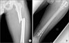
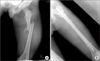 | Figure 1A 17-year-old male had a traffic accident. (A) Preoperative radiograph showing the shaft of the femur fracture. (B) Postoperative radiograph of the femur after intramedullary nailing.
|
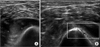
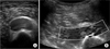 | Figure 2Ultrasonography of the left distal thigh showed an anechoic fluid collection like hematoma measuring 3.3×2.0×2.9 cm under the fasica of the vastus intermedius muscle (A) and turbulent blood flow in the hematoma (B).
|
 | Figure 3Fat suppressed coronal T1 (A) and axial T2 (B) magnetic resonance contrast images of the left distal thigh showed a pseudoaneurysm with internal hematoma which has heterogenous intensity signal under the vastus intermedius muscle. The margin of the hematoma was surrounded by a low intensity signal band shaped sac, and the tip of the distal interlocking screw was detected inside the hematoma. A, anterior; P, posterior; M, medial; L, lateral.
|
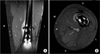
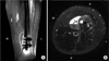
 | Figure 4Computed tomography angiography of the lower extremity showed a small vessel (arrow) around the distal screw area.
|
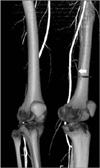
 | Figure 5An 18-year-old male had a traffic accident. (A) Preoperative radiograph showing the shaft of the femur fracture. (B) The radiograph of the femur taken six weeks after the operation showing callus formation at the fracture site.
|
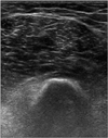
 | Figure 6Ultrasonography of the left distal thigh showed a hematoma measuring 4.3×8.0×1.3 cm (A) with turbulent blood flow (B).
|
 | Figure 7(A) Fat suppressed coronal T1 magnetic resonance (MR) contrast image of the left distal thigh showing a pseudoaneurysm with an internal hematoma. (B) Blood fluid level was shown adjacent to the distal interlocking screw tip in a hematoma on a fat suppressed axial T2 MR image. A, anterior; P, posterior; M, medial; L, lateral.
|
References
1. Cho SH, Kim DH, Jeong ST, et al. Therapeutic embolization for pseudoaneurysm of the anterior tibial artery after tibial nailing. J Korean Orthop Assoc. 2010; 45:238–242.

2. Bose D, Hauptfleisch J, McNally M. Delayed pseudoaneurysm caused by distal locking screw of a femoral intramedullary nail: a case report. J Orthop Trauma. 2006; 20:584–586.
3. Faure C, Merloz P. Transfixation: atlas of anatomical sections for the external fixation of limbs. Berlin, New York: Springer-Verlag;1987. p. 65–89.
4. Park SJ, Yang KH. Pseudoaneurysm of the superficial femoral artery following gamma nail fixation for trochanteric fracture: a case report. J Korean Orthop Assoc. 2000; 35:695–697.
5. Turek SL. Orthopaedics: principles and their application. 4th ed. Philadelphia: Lippincott;1984. p. 802–806.
6. Rutherford RB. Vascular surgery. 4th ed. Philadelphia: W.B. Saunders;1995. p. 1153–1161.
7. Han KJ, Won YY, Kim TY, Khang SY. Pseudoaneurysm of the anterior tibial artery after closed intramedullary nailing of a tibial shaft fracture: a case report. J Korean Orthop Assoc. 2002; 37:574–576.
8. Lee SH, Ahn JH. Diagnosis and treatment for delayed pseudoaneurysm of deep femoral artery: a case report. J Korean Orthop Assoc. 2008; 43:118–121.
9. Yoon HK, Kim BK, Shin DE, Kim MD, Chang JH. Pseudoaneurysm of superficial femoral artery following proximal femoral nail fixation. J Korean Fract Soc. 2004; 17:221–223.
10. Toursarkissian B, Allen BT, Petrinec D, et al. Spontaneous closure of selected iatrogenic pseudoaneurysms and arteriovenous fistulae. J Vasc Surg. 1997; 25:803–808.
 XML Download