

 PDF
PDF ePub
ePub Citation
 Print
Print


Abstract
Arteriovenous hemangiomas are found mainly in the limbs and scalp, and are common in women and children. However, there have been very limited reports concerning posttraumatic arteriovenous hemangioma, and none was reported in the medical literature regarding posttraumatic arteriovenous hemangioma of hypothenar eminence causing neurologic symptoms. We report a case of arteriovenous hemangioma of hypothenar eminence after recurrent trauma that was successfully treated by an excision of the mass and vein graft.
Figures and Tables
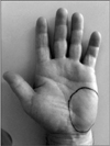
Figure 2
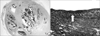
Color Doppler sonographic image showed a vascular mass including arterial blood flow of ulnar artery in the region of the hypothenar eminence (white arrow: ulnar artery).
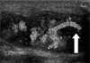
Figure 3
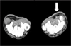
(A, B) Coronal T1 and T2-weighted magnetic resonance images show that homogeneous low signal intensity in hypothenar eminence of the left hand on T1-weighted image, which is converted to heterogenous high signal intensity at T2-weighted image. (C) Coronal T1-weighted enhanced magnetic resonance image shows well-defined enhancement and septum-like structures with low signal intensity of the hypothenar eminence. Magnetic resonance images show a vascular mass that is lobulated in the region of the hypothenar mass.
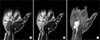
Figure 4
Intraoperative photograph showed a mass originated from the ulnar artery and compressed the ulnar nerve (white arrows: ulnar artery, right angled arrow: ulnar nerve).
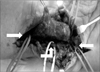
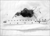
Figure 6
The deficit of the ulnar artery was grafted using dorsal metacarpal vein of the ipsilateral hand.
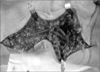
Figure 7
(A) Photomicrograph shows that the lesion is composed of an overgrowth of crowded blood vessels of various size and shape (H&E stain, ×10). (B) Elastic laminae and fibers (white arrow) in the arteries are clearly demonstrated (van Gieson stain, ×100).
References
1. Nazzi V, Messina G, Dones I, Ferroli P, Broggi G. Surgical removal of intramuscular arteriovenous hemangioma of the upper left forearm compressing radial nerve branches. J Neurosurg. 2008. 108:808–811.

2. Pulidori M, Capuano C, Mouchaty H, Cioffi F, Di Lorenzo N. Intramuscular thrombosed arteriovenous hemangioma of the upper right arm mimicking a neuroma of the ulnar nerve: case report. Neurosurgery. 2004. 54:770–771.
3. Acharya G, Merritt WH, Theogaraj SD. Hemangioendotheliomas of the hand: case reports. J Hand Surg Am. 1980. 5:181–182.
4. Enzinger FM, Weiss SW. Soft tissue tumors. 2001. 4th ed. St Louis: Mosby;837–887.
5. Legiehn GM, Heran MK. Venous malformations: classification, development, diagnosis, and interventional radiologic management. Radiol Clin North Am. 2008. 46:545–597.
6. Allen PW, Enzinger FM. Hemangioma of skeletal muscle. An analysis of 89 cases. Cancer. 1972. 29:8–22.
7. Cohen JM, Weinreb JC, Redman HC. Arteriovenous malformations of the extremities: MR imaging. Radiology. 1986. 158:475–479.
8. Perumal V, Francisco CJ. Arteriovenous hemangioma of the foot--a case report. Foot Ankle Surg. 2010. 16:e61–e62.
9. Pollard MA. Vascular tumors of the hand: a report of 3 cases. Hosp Physician. 2003. 3:57–61.
10. Hiroyasu N, Yukio S, Masayoshi U, Masahide I, Mitsunori O, Ryoji U. A case with arteriovenous hemangioma that occurred in the finger. Okinawa Med J. 2003. 41:62–64.
 XML Download