

 PDF
PDF ePub
ePub Citation
 Print
Print


Compression neuropathy of the ulnar nerve at the elbow is commonly caused by constriction in the fibrous cubital tunnel or an anatomical elbow deformity. However, many uncommon causes of compression have been described, including Osborne's band, ulnar nerve subluxation, anatomical confines of the cubital tunnel, elbow osteoarthritis, ganglia,1,2) a prominent medial head of the triceps,3) and an anconeus epitrochlearis.4) A ganglion can arise from either the synovium of joints or tendon sheaths or from tendons or nerves, and is filled with synovial fluid that may become jelly-like over time.1,2) Occasionally, one can cause problems, such as pain, numbness, and atrophy, especially when it compresses structures such as nerves. The presence of ganglia in the cubital tunnel leading to compression of the ulnar nerve and onset of symptoms has been previously described, although cases are rare. All cases of ganglia compressing the ulnar nerve have been shown to originate from the ulnohumeral joint and histological studies suggest that there is no "neural component." Specifically, all cases had a history of trauma to the elbow or showed degenerative arthritic findings on simple radiographs.1,2,5,6)
This report presents an extremely rare case of a fusiform epineural ganglion encompassing the ulnar nerve in the cubital tunnel with no connection to the elbow joint. In addition, this case showed no evidence of arthritic changes to the elbow joint and the preoperative range of motion was within normal limits. We obtained the patient's written informed consent for print and electronic publication of this case report.
CASE REPORT
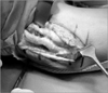
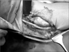
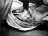
A 48-year-old man presented to our department with a 6-month history of progressive pain, numbness, and tingling around the medial epicondyle of the left elbow and the medial border of the forearm and hand. At the little finger, the two-point discrimination was 11 mm versus 6 mm on the right side. The patient had hand weakness with a decreased grip strength of 40 kg (right side, 50 kg) and pinch strength of 7 kg (right side, 19 kg). Froment's and Egawa's signs were positive, and there was atrophy of the adductor pollicis and first dorsal interosseous muscles. Electrophysiological studies demonstrated findings compatible with ulnar nerve compression (Table 1). At surgery, we found a fusiform epineural ganglion cyst (1.4 cm in diameter and 7.2 cm in length) encompassing the ulnar nerve in the cubital tunnel (Fig. 1). The proximal portion of the cyst was 1.4 cm superior to Struthers' ligament, and the distal portion extended into the right superior portion of Osborne's ligament; however, no connection to the elbow joint was found. The cyst contained a thick, jelly-like material. Under general anesthesia, we excised the ganglion cyst using microsurgery (Fig. 2) and performed ulnar nerve anterior transmuscular transposition (Fig. 3). Histology reported a "benign neural cyst with a myxoid matrix".
The patient was re-evaluated at 6 months postoperatively, both clinically and electrophysiologically. At 6 months, the patient was almost completely free of pain, tingling sensations, and numbness. In addition, his grip and pinch strength were improved and the adductor pollicis muscle wasting showed a dramatic recovery. A postoperative electrophysiological study showed some improvement, but the patient had not returned to normal at that time.
DISCUSSION
Our case has three distinct features. First, the fusiform epineural ganglion cyst had unique morphological characteristics. Specifically, it encompassed the ulnar nerve in the cubital tunnel. Second, the cyst did not originate from the medial aspect of the ulnohumeral joint. Finally, the clinical symptoms worsened gradually over a 6-month period, in contrast to reports of acute aggravation by a ganglion. Ganglia are not a common cause of peripheral nerve compression and have not been emphasized in the literature.1,2,5-10) Since the first report of a ganglion compressing the ulnar nerve in the hand,8) most studies have reported ganglia affecting the common peroneal nerve at the head of the fibula.7) Bowers and Doppelt2) reported that all ganglion cysts compressing the ulnar nerve in the cubital tunnel originated from the ulnohumeral joint and none were neural tissue. Ganglia commonly occur in middle-aged men, especially those with a history of elbow trauma.5,6) One case of ulnar nerve compression by a intraneural ganglion at the retrotrochlear groove was reported.5) That patient had symptoms for 3 months, no muscle atrophy was observed, and almost complete recovery of electrophysiological findings and clinical features were observed after ganglion excision. Boursinos and Dimitriou7) reported a patient who developed muscle wasting due to a longer symptomatic period; the ganglion was 2 cm in diameter, originated from the epineurium without being surrounded by nerve fibers, and had no connection to the elbow joint. In our case, the 6-month history of progressive symptoms in addition to the ganglion encompassing the nerve may explain the lack of complete recovery of electrophysiological parameters despite a significant clinical improvement 6 months postoperatively. Since ganglia are not a common cause of cubital tunnel syndrome, they are often misdiagnosed because preoperative imaging studies are not always performed. Since palpation before surgery revealed no evidence suggesting a mass in the cubital tunnel and the symptoms and physical examination made us suspect typical compressive neuropathy of the cubital tunnel, neither ultrasound (US) nor magnetic resonance imaging (MRI) was performed. MRI provides the best view of the cyst contents and its relationship to adjacent neurovascular structures. In addition, computed tomography and US may be useful. However, the patient's history, clinical symptoms, and electrophysiological findings are also sufficient for diagnosing typical compressive neuropathy. Total excision of the epineural ganglion may not always be possible without damaging the nerve, especially if the ganglion is intraneural.1-4) In all reported cases, an early diagnosis and careful excision was associated with a satisfactory outcome.5,6,9,10) Due to removed intermuscular septum, Osborne's ligament, flexor-pronator fascia during the exploration and excision for the ganglion, anterior transposition of the ulnar nerve was considered, as means of decompression and protection. As in other reports, the fusiform epineural ganglion was excised and ulnar nerve anterior transmuscular transposition, which the author prefers, was performed. At the 6-month follow-up, the patient was almost completely free of pain, sensory impairment, and numbness. Additionally, grip and pinch strength were improved and the adductor pollicis muscle wasting showed a dramatic recovery.

 XML Download