

 PDF
PDF ePub
ePub Citation
 Print
Print


Parsonage-Turner syndrome (PTS) can be a problem for both patients and physicians. PTS patients typically present with sudden onset of arm pain followed by muscle atrophy of the shoulder girdle and arm, which induces upper extremity weakness.1) Treatment of PTS is usually supportive and involves a combination of analgesics, immobilization and physical therapy, but some degree of sequelae is reported in about 10% of patients.2,3) Steroid injection is reported to show better treatment results than others. In previous studies, early corticosteroid therapy might have positively influenced the acute pain phase in some patients; however, the use of steroids is controversial in the muscle weakness phase.
This article presents cases of PTS with steroid therapy in the muscle weakness phase. This study has a small study group of six cases, but is the first one on the effects of steroids after the onset of muscle weakness, showing better treatment results when steroids were injected during the weakness phase of PTS.
CASE REPORT
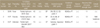
Group 1 had three cases. The mean age was 48 years old, ranging from 36 to 61. Case 1 had a mild fever history, and case 2 had a trauma history of sudden crepitus following severe burning pain at the shoulder and arm during work. All visited other medical institutions and were prescribed medication but had little pain relief, with pain continuing again after a mean of 23 days (range 17-27) from the onset of symptoms. The medical records of the other medical institutions did not report numbness at the time of pain onset. Patients were transferred to our hospital with a chief complaint of muscle weakness of the upper extremities at about one month from the onset of pain. On physical examination, muscle power weakness was about Medical Research Council (MRC) grade 3. All range of motion from the shoulder joint to finger joints was checked and a mild degree of muscle atrophy was observed at the deltoid, biceps, and triceps muscles. Range of motion was normal, and a spurling test of the neck was negative. Paresthesia of the upper limbs was not checked. Specific findings were not checked by magnetic resonance imaging, except for mild disc herniation of cervical level 4/5, 5/6. Electromyogram (EMG) found lesions at the area of the brachial plexus or cervical plexus. Based on the clinical course and the EMG findings, conservative treatment such as physical therapy including muscle-straightening exercises, were performed with a diagnosis of PTS. Recovery parameters were set by muscle strength, timing of recovery, and Rankin score. For recovery parameters, recovery of muscle strength was defined by strength of more than 80% power compared to the normal side. Improvement of muscle power was observed after 8 months (MRC grade 4), and patients were able to work normally after 12 months (grade 5) (Table 3). Summarized data for Group 1 are in Table 1.
Group 2 had three cases with a mean age of 47 years old, ranging from 39 to 51. Patients were engaged in an occupation that used the arms frequently, and none had a specific trauma or disease history. Two patients had visited other medical institutions with a chief complaint of burning pain of the upper extremity and pharmacological treatment was given for pain control although this gave no pain relief. The patients visited our hospital within 14 days of pain onset. In one case, the patient visited our hospital just after pain onset because of pain in the left upper extremity and shoulder. At the time of the visit, no sign of motor or sensory weakness of the affected upper extremities was observed. On physical examination at admission, the range of motion of the upper limbs was normal, and tenderness and tinel sign were positive at the supraclavicular brachial plexus, but muscle atrophy was not found. Muscle power, sensation, and tendon reflex were all normal. A spurling test of the neck was negative, and various tests for shoulder lesions such as rotator cuff tear were all negative. Simple X-ray of the neck and shoulder joint showed no abnormal findings. After 5 weeks from onset, pain was relieved but weakness of muscle power at left upper limb appeared, with grade 3 for elbow flexion. EMG performed at that time found a lesion at the area of the left brachial plexus. Based on the clinical course and EMG findings, the patient was diagnosed with PTS. A dose regimen of steroids followed, with a two week course of oral prednisolone, 60 mg daily in the first week, and tapering to the dosage by 10 mg per day with a 5 mg as the last step. Oral prednisolone was accompanied with physical therapy within 7 days of the onset of muscle weakness (one case just after onset, two cases within 5 days of onset). No side effects of steroid treatment were observed. Recovery parameters were as for Group 1. After 8 months, muscle strength was restored (MRC grade 5) and the patient was able to return to his work (Table 3). The summarized data of Group 2 is described in Table 2.
DISCUSSION
PTS was first reported by Freinberg in 1897, but the disease name was not established until Personage and Turner described a series of 136 clinical cases in 1948. Other terms used to describe this disease entity include neuralgic amyotrophy, brachial plexus neuritis, idiopathic brachial plexopathy, acute brachial radiculitis.1,3) However, the general term Parsonage-Turner syndrome is most commonly used. The etiology of this condition remains unclear, but an immune attack on the brachial plexus or its braches within the limb triggered by various preceding events, such as infection, vaccination, pregnancy and parturition, surgery, radiation, intravenous heroin use, or treatment with interferon, has been suggested as a cause, although pathological evidence is scant. Trauma is not a certain risk factor, but is recognized as a contributing factor after Mulvey et al2) reported that mild trauma could induce symptoms of neuritis.3) One case in group 1 was associated with trauma, and the others were related to mild infection. Symptoms of PTS vary widely, but the typical clinical course starts with acute, severe, aching, unilateral shoulder and proximal arm pain lasting from days to a few weeks. When the pain abates, painless shoulder girdle and arm weakness develops, more prominent in the upper plexus muscles including deltoid, supra or infraspinatus, biceps. Forearm and hand muscles are less frequently involved.4-6) For numbness, different viewpoints exists; Tsairis et al7) reported 67% of cases with numbness, while Misamore and Lehman5) reported that no cases with numbness.
Treatment is usually supportive and involves a combination of analgesics, immobilization and physical therapy.1,3,5,6) Recently, steroid injection was reported to give better treatment results, however, these reports were based on limited resources such as a few case reports so controversy remains about the effects of the treatment.6,8) Non-randomized studies provide some evidence to support shortening the time of intense pain and hastening motor nerve recovery when the corticosteroids are administered during the acute phase of the condition.9) Anecdotal evidence suggests that their use leads to a more rapid resolution of the painful phase of the illness, in particular when used early in its course, although they does not seem to influence the ultimate prognosis. Non-controlled clinical observations suggest that very early treatment with corticosteroids in some cases results in prompt pain resolution, with no or minimal weakness.10) In this study, patients of group 1 returned to work after 6 months from the onset of pain, and worked normally after 12 months. However, in group 2, muscle strength was restored after 8 months from the onset of the pain, with return to normal work within 12 months (p=0.001) (Table 3). However, statistical comparisons of steroidtreated patients with the untreated group showed no differences in baseline variables such as age, sex, preceding history and the occurrence of atrophy, or weakness symptoms during the attack.
No study has reported on the efficacy of steroid treatment in the muscle weakness phase. Although this report lacks a large number of cases and a follow-up period, it is meaningful as the first study on the efficacy of steroid injection in the muscle weakness phase of PTS. We propose that even in this phase, steroid injection is possible to improve recovery time and enable rapid return to daily life.

 XML Download