

 PDF
PDF ePub
ePub Citation
 Print
Print


Abstract
Purpose
This study examined the clinical efficacy of an anterior cervical discectomy and fusion (ACDF) with PEEK (polyetheretherketone) cage alone with regard to the clinical and radiological outcomes, as well as the risk factors for the cage subsidence.
Materials and Methods
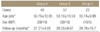
A total of 128 patients who underwent group A (1-level, n=48), group B (2-levels, n=57), group C (3-levels, n=23) ACDF using a PEEK cage alone were enrolled in this study. The fusion rate, segmental kyphosis were assessed by radiographs. The clinical outcomes were assessed using the Visual Analog Scale (VAS) and Neck Disability Index (NDI). The risk factors for cage subsidence were analyzed according to the difference in incidence between the subsidence group and non-subsidence group.
Results
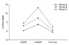
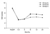
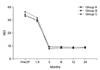
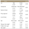
Solid fusion was achieved in 93.8% (45/48), 71.9% (41/57) and 69.6% (15/23) of subjects in group A, B and C, respectively. Segmental kyphosis was observed in 22.9% (11/48), 43.9% (25/57) and 47.8% (11/23) of subjects in group A, B and C, respectively. The VAS scores changed from 7.79±1.01 in group A, 7.74±1.09 in group B, 7.91±0.79 in group C preoperatively to 4.23±1.29 in group A, 5.25±1.34 in group B and 5.35±1.07 in group C at the last follow up. In addition, the NDI was also improved at the last follow up. The VAS score and NDI at the last follow up were similar in the subsidence and non-subsidence group. The 3-level ACDF (p=0.05), osteoporosis (p=0.01), and old age (p=0.01) were the risk factors for cage subsidence.
Conclusion
Only 1 level ACDF with PEEK cage alone was similar in clinical and radiologic (solid fusion rate, local kyphosis) outcomes compared to ACDF with published other modalities. Old age, 3 fusion level, osteoporosis, and C6-7 fusion were risk factors for the cage subsidence with higher complication rates.
Figures and Tables
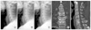 | Figure 1A 52-year-old male status post ACDF with PEEK (Solis) cage alone at C6-C7, (A) postoperative plain lateral radiograph; (B) plain lateral radiograph 1.5 months postsurgery showing bone bridging; (C) plain lateral radiograph 2 years postsurgery showing normal alignment, no collapse and good consolidation of bone graft in the cage; (D) coronal CT image at 2 years postsurgery showing bony bridging (connection) and definite consolidation of graft-bone in the cage; (E) sagittal CT image at 2 years postsurgery.
|
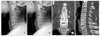 | Figure 3A 47-year-old male status post ACDF with PEEK (Solis) cage alone at C4-C5, 5-6, 6-7, (A) postoperative plain lateral radiograph; (B) plain lateral radiograph 2 years postsurgery showing cage subsidence and no bony consolidation of bone graft in the cage; (C) coronal CT image at 2 years postsurgery and (D) sagittal CT image at 2 years postsurgery showing no bony bridging (connection) in the graft-bone, and a vacant space in the cage (no bony tissue).
|


References
1. Robinson RA, Smith GW. Anterolateral cervical disc removal and interbody fusion for cervical disc syndrome. Bull Johns Hopkins Hosp. 1955. 96:223–224.

2. Liao JC, Niu CC, Chen WJ, Chen LH. Polyetheretherketone (PEEK) cage filled with cancellous allograft in anterior cervical discectomy and fusion. Int Orthop. 2008. 32:643–648.
3. Cho DY, Lee WY, Sheu PC. Treatment of multilevel cervical fusion with cages. Surg Neurol. 2004. 62:378–385.
4. Cho DY, Liau WR, Lee WY, Liu JT, Chiu CL, Sheu PC. Preliminary experience using a polyetheretherketone (PEEK) cage in the treatment of cervical disc disease. Neurosurgery. 2002. 51:1343–1349.
5. Gercek E, Arlet V, Delisle J, Marchesi D. Subsidence of stand-alone cervical cages in anterior interbody fusion: warning. Eur Spine J. 2003. 12:513–516.
6. Song KJ, Taghavi CE, Lee KB, Song JH, Eun JP. The efficacy of plate construct augmentation versus cage alone in anterior cervical fusion. Spine (Phila Pa 1976). 2009. 34:2886–2892.
7. Song KJ, Taghavi CE, Hsu MS, Lee KB, Kim GH, Song JH. Plate augmentation in anterior cervical discectomy and fusion with cage for degenerative cervical spinal disorders. Eur Spine J. 2010. 19:1677–1683.
8. Kandziora F, Pflugmacher R, Scholz M, et al. Treatment of traumatic cervical spine instability with interbody fusion cages: a prospective controlled study with a 2-year follow-up. Injury. 2005. 36:Suppl 2. B27–B35.
9. Banwart JC, Asher MA, Hassanein RS. Iliac crest bone graft harvest donor site morbidity. A statistical evaluation. Spine (Phila Pa 1976). 1995. 20:1055–1060.
10. Sawin PD, Traynelis VC, Menezes AH. A comparative analysis of fusion rates and donor-site morbidity for autogeneic rib and iliac crest bone grafts in posterior cervical fusions. J Neurosurg. 1998. 88:255–265.
11. Floyd T, Ohnmeiss D. A meta-analysis of autograft versus allograft in anterior cervical fusion. Eur Spine J. 2000. 9:398–403.
12. Connolly PJ, Esses SI, Kostuik JP. Anterior cervical fusion: outcome analysis of patients fused with and without anterior cervical plates. J Spinal Disord. 1996. 9:202–206.
13. Bose B. Anterior cervical instrumentation enhances fusion rates in multilevel reconstruction in smokers. J Spinal Disord. 2001. 14:3–9.
14. Lowery GL, McDonough RF. The significance of hardware failure in anterior cervical plate fixation. Patients with 2- to 7-year follow-up. Spine (Phila Pa 1976). 1998. 23:181–186.
15. Fujibayashi S, Shikata J, Kamiya N, Tanaka C. Missing anterior cervical plate and screws: a case report. Spine (Phila Pa 1976). 2000. 25:2258–2261.
16. Vavruch L, Hedlund R, Javid D, Leszniewski W, Shalabi A. A prospective randomized comparison between the cloward procedure and a carbon fiber cage in the cervical spine: a clinical and radiologic study. Spine (Phila Pa 1976). 2002. 27:1694–1701.
17. Chou YC, Chen DC, Hsieh WA, et al. Efficacy of anterior cervical fusion: comparison of titanium cages, polyetheretherketone (PEEK) cages and autogenous bone grafts. J Clin Neurosci. 2008. 15:1240–1245.
18. Kulkarni AG, Hee HT, Wong HK. Solis cage (PEEK) for anterior cervical fusion: preliminary radiological results with emphasis on fusion and subsidence. Spine J. 2007. 7:205–209.
19. Lim TH, Kwon H, Jeon CH, et al. Effect of endplate conditions and bone mineral density on the compressive strength of the graft-endplate interface in anterior cervical spine fusion. Spine (Phila Pa 1976). 2001. 26:951–956.
20. Thomé C, Krauss JK, Zevgaridis D. A prospective clinical comparison of rectangular titanium cages and iliac crest autografts in anterior cervical discectomy and fusion. Neurosurg Rev. 2004. 27:34–41.
21. Schmieder K, Wolzik-Grossmann M, Pechlivanis I, Engelhardt M, Scholz M, Harders A. Subsidence of the wing titanium cage after anterior cervical interbody fusion: 2-year follow-up study. J Neurosurg Spine. 2006. 4:447–453.
22. Wilke HJ, Kettler A, Goetz C, Claes L. Subsidence resulting from simulated postoperative neck movements: an in vitro investigation with a new cervical fusion cage. Spine (Phila Pa 1976). 2000. 25:2762–2770.
23. Oxland TR, Lund T. Biomechanics of stand-alone cages and cages in combination with posterior fixation: a literature review. Eur Spine J. 2000. 9:Suppl 1. S95–S101.
24. Goh JC, Wong HK, Thambyah A, Yu CS. Influence of PLIF cage size on lumbar spine stability. Spine (Phila Pa 1976). 2000. 25:35–39.
25. Truumees E, Demetropoulos CK, Yang KH, Herkowitz HN. Failure of human cervical endplates: a cadaveric experimental model. Spine (Phila Pa 1976). 2003. 28:2204–2208.
 XML Download