

 PDF
PDF ePub
ePub Citation
 Print
Print


Abstract
Secondary osteosarcoma has a relatively higher incidence in middle aged persons than in children. Radiation-induced osteosarcoma occurs in approximately 1% of patients who have been treated with more than 2,500 cGy. The time interval from radiation to onset of secondary osteosarcoma is approximately 10 to 15 years. A 51-year-old female who have been treated with radiation for angiomyxoma was hospitalized due to right hip pain. She had a minor trauma 2 weeks prior to hospitalization. A day before hospitalization, she experienced a second trauma by fall, and then, severe hip pain developed. A radiograph of the patient showed femoral neck fracture with sclerotic change of fractured margin. We diagnosed the patient as having a neglected femoral neck fracture and treated it with closed reduction using cannulated screw fixation. At 6 months post-surgery, the patient had residual pain of the right hip and we could find overproduced callus at the fracture site. Through further evaluation, we diagnosed this as secondary osteosarcoma with pulmonary metastasis. We report this case to make a warning about a misdiagnosed osteosarcoma as a simple femoral neck fracture.
Figures and Tables
Figure 1
Initial anteroposterior radiograph shows a transcervical femoral neck fracture with modest displacement in Garden type 3. Sclerotic change is seen at the fracture site. No periosteal reaction is found and any other findings looked like mass are not observed.
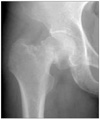
Figure 2
Postoperative radiograph (A) and radiograph made 4 months after surgery (B) show internal fixation with three cannulated screws at right femoral neck. In the radiograph made 4 months after surgery(B), no evidence of union is found. Blurring mixed with calcification around the fracture site is seen. Anteroposterior radiograph made 6 months after surgery (C) shows overpruded callus. There is loss of cortical and cancellous bony architecture with destructive and heterogenous shadow.
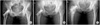
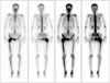
Figure 3
Bone scintigraphy images show asymmetrical intensed increased uptake in right femoral head, neck and proximal portion.
References
1. Kalra S, Grimer RJ, Spooner D, Carter SR, Tillman RM, Abudu A. Radiation-induced sarcomas of bone: factors that affect outcome. J Bone Joint Surg Br. 2007. 89:808–813.
2. Koshurnikova NA, Gilbert ES, Sokolnikov M, et al. Bone cancers in Mayak workers. Radiat Res. 2000. 154:237–245.

3. Bielack SS, Kempf-Bielack B, Delling G, et al. Prognostic factors in high-grade osteosarcoma of the extremities or trunk: an analysis of 1,702 patients treated on neoadjuvant cooperative osteosarcoma study group protocols. J Clin Oncol. 2002. 20:776–790.
4. Souhami RL, Craft AW, Van der Eijken JW, et al. Randomised trial of two regimens of chemotherapy in operable osteosarcoma: a study of the European Osteosarcoma Intergroup. Lancet. 1997. 350:911–917.
5. Grimer RJ, Cannon SR, Taminiau AM, et al. Osteosarcoma over the age of forty. Eur J Cancer. 2003. 39:157–163.
6. Shaheen M, Deheshi BM, Riad S, et al. Prognosis of radiation-induced bone sarcoma is similar to primary osteosarcoma. Clin Orthop Relat Res. 2006. 450:76–81.
7. Okada K, Hasegawa T, Nishida J, et al. Osteosarcomas after the age of 50: a clinicopathologic study of 64 cases-an experience in northern Japan. Ann Surg Oncol. 2004. 11:998–1004.
8. Jeon DG, Lee SY, Cho WH, Song WS, Park JH. Primary osteosarcoma in patients older than 40 years of age. J Korean Med Sci. 2006. 21:715–718.
9. Sadoghi P, Leithner A, Clar H, et al. The threat of misdiagnosis of primary osteosarcoma over the age of 60: a series of seven cases and review of the literature. Arch Orthop Trauma Surg. 2010. 130:1251–1256.
10. Sass M, Melcher I, Schaser KD, Stropahl G, Mittlmeier T. Management problems in the diagnostic work-up and treatment of pathological fracture of the proximal femur: clinical case report. Unfallchirurg. 2006. 109:251–255.
 XML Download