

 PDF
PDF ePub
ePub Citation
 Print
Print


Abstract
Peroneus quartus tendon is one of a group of accessory peroneal tendons present in humans. Associated pathologies include an attritional tear in the peroneus tendon, stenosing tendinitis, and peroneal tendon subluxation or dislocation. A 22-year old female patient, who presented suffering with lateral ankle pain and instability after ankle sprain, visited this hospital. The physical examination and MRI indicated a longitudinal tear of peroneus brevis tendon or chronic stenosing tendinitis caused by peroneus quartus tendon. Tenosynovectomy and an excision of the peroneus quartus tendon were performed. Satisfactory results were obtained at the 1 year follow-up after surgery. Orthopedic surgeons should consider the possible presence of the peroneus quartus tendon in a differential diagnosis of chronic lateral ankle pain.
Figures and Tables
Figure 1
Drawing illustrates a peroneus quartus tendon (arrowhead) located posterior to the peroneus brevis (long arrow) and peroneus longus (short arrow) tendons. Its most common insertion site is the retrotrochlear eminence of calcaneus (curved arrow).

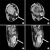
Figure 2
Sequential axial MR images show an accessory tendon (arrowhead) behind peroneus longus (long arrow) and peroneus brevis (short arrow) tendon. (A, B) This accessory tendon identified with distinct tendon slip within the muscle belly is a peroneus quartus tendon simulating a torn peroneus brevis tendon. (C, D) A peroneus quartus tendon (arrowhead) located close to its insertion into retrotrochlear eminence (curved arrow).


References
1. Zammit J, Singh D. The peroneus quartus muscle. Anatomy and clinical relevance. J Bone Joint Surg Br. 2003. 85:1134–1137.
2. Hecker P. Study of the peroneus of the tarsus. Anat Rec. 1923. 26:79–82.
3. Sobel M, Levy ME, Bohne WH. Congenital variations of the peroneus quartus muscle: an anatomic study. Foot Ankle. 1990. 11:81–89.

4. Trono M, Tueche S, Quintart C, Libotte M, Baillon J. Peroneus quartus muscle: a case report and review of the literature. Foot Ankle Int. 1999. 20:659–662.
5. Regan TP, Hughston JC. Chronic ankle sprain secondary to anormalous peroneal tendon: a case report. Clin Orthop Relat Res. 1977. 123:52–54.
6. Buschmann WR, Cheung Y, Jahss MH. Magnetic resonance imaging of anormalous leg muscles: accessory soleus, peroneus quartus and the flexor digitorum longus accessories. Foot Ankle. 1991. 12:109–116.
7. Mick CA, Lynch F. Reconstruction of the peroneal retinaculum using the peroneus quartus. A case report. J Bone Joint Surg Am. 1987. 69:296–297.
8. Sammarco GJ, Diraimondo CV. Chronic peroneus brevis tendon lesions. Foot Ankle. 1989. 9:163–170.
9. Sobel M, Geppert MJ, Warren RF. Chronic ankle instability as a cause of peroneal tendon injury. Clin Orthop Relat Res. 1993. 296:187–191.
10. Sobel M, Geppert MJ, Olson EJ, Bohne WH, Arnoczky SP. The dynamics of peroneus brevis tendon splits: a proposed mechanism,technique of diagnosis, and classification of injury. Foot Ankle Int. 1992. 13:413–422.
 XML Download