

 PDF
PDF ePub
ePub Citation
 Print
Print


Abstract
Purpose
To analyze, using finite element model analysis, the causes of postoperative pain in patients who had arthroscopic treatment for femoroacetabular impingement (FAI).
Materials and Methods
Ten patients with FAI treated by arthroscopic surgery between July 2004 and July 2007 were selected. Five cases whose condition improved to a pain score of 3 postoperatively were assigned to comparative group A and 5 cases who had a second operation done due to a pain score of 1 were assigned to experimental group B. Finite element model analysis was done for the impingement test position. Femoral offset and alpha angle were measured to compare with contact pressure or von Mises stress.
Figures and Tables
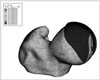 | Figure 1Three dimensional reconstructive view of femoral head. The blue colored covering of femoral head shows the contact area when hip joint rotates, and the sequential color table on the left side indicates the degree of contact pressure and von Mises stress.
|
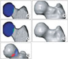
 | Figure 2The correlation of motion and pressure was interpreted by the flexion degree (Z axis) and the internal rotation degree (X axis).
|
 | Figure 3(A, B) Preoperative finite element model of a patient in group B. A small portion of femoral head neck junction had an increased contact pressure which indicates a clinical "bump". (C, D) Postoperative model shows that the "bump" was not removed sufficiently, and the contact pressure increased. (E) This model shows the removed bump (striped diagram) and portions of concentrated stress (filled diagram). This figure shows that the concentrated stress portion had not been removed successfully.
|
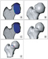 | Figure 4(A, B) Preoperative finite element model of a patient in group A. A small portion of femoral head neck junction had an increased contact pressure which indicates a clinical "bump". (C, D) Postoperative model shows that the "bump" was removed sufficiently, and the contact pressure was 0 at the maximal internal rotation degree. (E) This model shows removed bump (striped diagram) and portions of concentrated stress (filled diagram). This figure shows that the concentrated stress portion had been removed successfully.
|
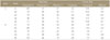
Table 1
The von Mises Stress and Contact Pressure of Groups A and B (Group A, Postoperative Pain Score was 3.; Group B, Postoperative Pain Score was 1.)
![]()

References
1. Philippon MJ, Schenker ML, Briggs KK, Kuppersmith DA, Maxwell RB, Stubbs AJ. Revision hip arthroscopy. Am J Sports Med. 2007. 35:1918–1921.

2. Robertson WJ, Kadrmas WR, Kelly BT. Arthroscopic management of labral tears in the hip: a systematic review of the literature. Clin Orthop Relat Res. 2007. 455:88–92.
3. Heyworth BE, Shindle MK, Voos JE, Rudzki JR, Kelly BT. Radiologic and intraoperative findings in revision hip arthroscopy. Arthroscopy. 2007. 23:1295–1302.
4. Outerbridge RE. The etiology of chondromalacia patellae. J Bone Joint Surg Br. 1961. 43:B:752–B:757.
5. Shima Y. Standard for evaluation of osteoarthritis of the hip. J Jpn Orthop Assoc. 1971. 45:813–833.
6. Moglo KE, Shirazi-Adl A. On the coupling between anterior and posterior cruciate ligaments, and knee joint response under anterior femoral drawer in flexion: a finite element study. Clin Biomech (Bristol, Avon). 2003. 18:751–759.
7. Chegini S, Beck M, Ferguson SJ. The effects of impingement and dysplasia on stress distributions in the hip joint during sitting and walking: a finite element analysis. J Orthop Res. 2009. 27:195–201.
8. Vulpius O, Stöffel A. Orthopaadische operationslehre. 1913. Stuttgart, Germany F. Enke.
9. Ganz R, Parvizi J, Beck M, Leunig M, Nötzli H, Siebenrock KA. Femoroacetabular impingement: a cause for osteoarthritis of the hip. Clin Orthop Relat Res. 2003. 417:112–120.
10. Lavigne M, Parvizi J, Beck M, Siebenrock KA, Ganz R, Leunig M. Anterior femoroacetabular impingement: part I. Techniques of joint preserving surgery. Clin Orthop Relat Res. 2004. 418:61–66.
11. Elmslie RC. Remarks on aetiological factors in osteoarthritis of the hip-joint. BMJ. 1933. 1:1–3.
12. Goodman DA, Feighan JE, Smith AD, Latimer B, Buly RL, Cooperman DR. Subclinical slipped capital femoral epiphysis. Relationship to osteoarthrosis of the hip. J Bone Joint Surg Am. 1997. 79:1489–1497.
13. Murray RO. The aetiology of primary osteoarthritis of the hip. Br J Radiol. 1965. 38:810–824.
14. Solomon L. Patterns of osteoarthritis of the hip. J Bone Joint Surg Br. 1976. 58:176–183.
15. Tönnis D, Heinecke A. Acetabular and femoral anteversion: relationship with osteoarthritis of the hip. J Bone Joint Surg Am. 1999. 81:1747–1770.
16. Law WA. Osteoarthritis of the hip. 1952. London: Butterworth & Co Ltd..
17. Stulberg SD, Cordell LD, Harris WH, Ramsey PL, MacEwen GD. Unrecognized childhood hip disease: a major cause of idiopathic osteoarthritis of the hip. The Hip. Proc 3rd meeting of The Hip Society. 1975. St Louis: CV Mosby Co.;212–228.
18. Ito K, Minka MA 2nd, Leunig M, Werlen S, Ganz R. Femoroacetabular impingement and the cam-effect. A MRI-based quantitative anatomical study of the femoral head-neck offset. J Bone Joint Surg Br. 2001. 83:171–176.
19. Nötzli HP, Wyss TF, Stoecklin CH, Schmid MR, Treiber K, Hodler J. The contour of the femoral head-neck junction as a predictor for the risk of anterior impingement. J Bone Joint Surg Br. 2002. 84:556–560.
20. Philippon MJ, Briggs KK, Yen YM, Kuppersmith DA. Outcomes following hip arthroscopy for femoroacetabular impingement with associated chondrolabral dysfunction: minimum two-year follow-up. J Bone Joint Surg Br. 2009. 91:16–23.
21. Hwang DS, Kang C, Cha SM, Kim JH. Second-look hip arthroscopy after arthroscopic labrectomy of the hip: preliminary report. J Korean Orthop Assoc. 2009. 44:480–485.
22. Wenger DE, Kendell KR, Miner MR, Trousdale RT. Acetabular labral tears rarely occur in the absence of bony abnormalities. Clin Orthop Relat Res. 2004. 426:145–150.
23. Bachtar F, Chen X, Hisada T. Finite element contact analysis of the hip joint. Med Biol Eng Comput. 2006. 44:643–651.
24. Russell ME, Shivanna KH, Grosland NM, Pedersen DR. Cartilage contact pressure elevations in dysplastic hips: a chronic overload model. J Orthop Surg Res. 2006. 1:6.
25. Murphy SB, Ganz R, Müller ME. The prognosis in untreated dysplasia of the hip. A study of radiographic factors that predict
the outcome. J Bone Joint Surg Am. 1995. 77:985–989.
26. Mardones RM, Gonzalez C, Chen Q, Zobitz M, Kaufman KR, Trousdale RT. Surgical treatment of femoroacetabular impingement: evaluation of the effect of the size of the resection. J Bone Joint Surg Am. 2005. 87:273–279.
27. Beck M, Kalhor M, Leunig M, Ganz R. Hip morphology influences the pattern of damage to the acetabular cartilage: femoroacetabular impingement as a cause of early osteoarthritis of the hip. J Bone Joint Surg Br. 2005. 87:1012–1018.
28. Bizzini M, Notzli HP, Maffiuletti NA. Femoroacetabular impingement in professional ice hockey players: a case series of 5 athletes after open surgical decompression of the hip. Am J Sports Med. 2007. 35:1955–1959.
29. Mardones R, Lara J, Donndorff A. Surgical correction of "cam-type" femoroacetabular impingement: a cadaveric comparison
of open versus arthroscopic debridement. Arthroscopy. 2009. 25:175–182.
 XML Download