

 PDF
PDF ePub
ePub Citation
 Print
Print


Abstract
Purpose
The purpose of this study is to evaluate the clinical and radiological outcomes of proximal, middle and distal third humeral fractures treated with the minimally invasive plate osteosynthesis (MIPO).
Materials and Methods
Thirty-one patients with the proximal, middle or distal third humeral fractures underwent MIPO. There were 12 men and 19 women with an average age of 46.7 years. The radiological outcomes of bony union and anatomical reduction were evaluated. The clinical outcomes were assessed by measuring the range of shoulder and elbow motion, UCLA scores, KSS sores and the postoperative complications.
Results
Fracture union was obtained in all patients at an average of 18.4 weeks. According to the UCLA scores, 9 were excellent and 3 were good for the proximal humeral fractures. For the middle and distal third fractures, UCLA scoring system showed excellent results in 15 cases and good results in 4 cases. The average KSS scores of proximal and shaft fracture were 92.5 and 98.6, respectively. Complications developed in 3 patients: one had radial nerve palsy, one had a fracture adjacent to the plate distally, and one had a rotational deformity.
Figures and Tables
Figure 1
The axillary nerve is palpated and protected by index finger while the plate is inserted to the proximal humerus fracture site through the deltoid splitting incision.
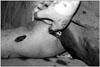
Figure 2
A K-wire is inserted to the proximal humerus through the plate and reduction is achieved by purchasing the cortical screw to the humeral shaft.
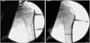
Figure 3
A 4.5 mm narrow LCP plate is placed on the anterior aspect of the arm under the C-arm so as to draw two longitudinal lines of 3 cm on proximal and distal portion, respectively.
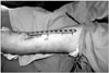
Figure 4
Preoperative AP and lateral radiographs of a 28-year-old male show B2 fracture of humerus shaft, clavicle shaft fracture and scapular body fracture. Postoperative radiographs at 10 months show union of the fracture with good alignment.

Figure 5
(A) A 3.5/4.5 mm metaphyseal LCP plate is molded to adapt it to the anterior surface of lateral column of distal humerus which is concave and inclined anteriorly in shape. (B) A 24 year-old female with distal third humeral fracture was treated with minimally invasive procedure. (C) Postoperative radiographs at 6 months showed a union of the fracture.

References
1. Volgas DA, Stannard JP, Alonso JE. Nonunions of the humerus. Clin Orthop Relat Res. 2004. 419:46–50.

2. Mills HJ, Horne G. Fractures of the proximal humerus in adults. J Trauma. 1985. 25:801–805.
3. Sarmiento A, Kinman PB, Galvin EG, Schmitt RH, Phillips JG. Functional bracing of fractures of the shaft of the humerus. J Bone Joint Surg Am. 1977. 59:596–601.
4. Flinkkilä T, Hyvönen P, Lakovaara M, Linden T, Ristiniemi J, Hämäläinen M. Intramedullary nailing of humeral shaft fractures. A retrospective study of 126 cases. Acta Orthop Scand. 1999. 70:133–136.
5. Rommens PM, Blum J, Runkel M. Retrograde nailing of humeral shaft fractures. Clin Orthop Relat Res. 1998. 350:26–39.
6. Apivatthakakul T, Patiyasikan S, Luevitoonvechkit S. Danger zone for locking screw placement in minimally invasive plate osteosynthesis (MIPO) of humeral shaft fractures: a cadaveric study. Injury. 2010. 41:169–172.
7. Zhiquan A, Bingfang Z, Yeming W, Chi Z, Peiyan H. Minimally invasive plating osteosynthesis (MIPO) of middle and distal third humeral shaft fractures. J Orthop Trauma. 2007. 21:628–633.
8. Livani B, Belangero WD. Bridging plate osteosynthesis of humeral shaft fractures. Injury. 2004. 35:587–595.
9. Ji F, Tong D, Tang H, et al. Minimally invasive percutaneous plate osteosynthesis (MIPPO) technique applied in the treatment of humeral shaft distal fractures through a lateral approach. Int Orthop. 2009. 33:543–547.
10. Paavolainen P, Björkenheim JM, Slätis P, Paukku P. Operative treatment of severe proximal humeral fractures. Acta Orthop Scand. 1983. 54:374–379.
11. Shin SI, Song KW, Lee JY, et al. Treatment of two- and three-part fracture of proximal humerus using LCP. J Korean Shoulder Elbow Soc. 2007. 10:204–211.
12. Vander Griend R, Tomasin J, Ward EF. Open reduction and internal fixation of humeral shaft fractures. Results using AO plating techniques. J Bone Joint Surg Am. 1986. 68:430–433.
13. Farouk O, Krettek C, Miclau T, Schandelmaier P, Guy P, Tscherne H. Minimally invasive plate osteosynthesis and vascularity: preliminary results of a cadaver injection study. Injury. 1997. 28:Suppl 1. A7–A12.
14. Rouleau DM, Laflamme GY, Berry GK, Harvey EJ, Delisle J, Girard J. Proximal humerus fractures treated by percutaneous locking plate internal fixation. Orthop Traumatol Surg Res. 2009. 95:56–62.
15. Apivatthakakul T, Arpornchayanon O, Bavornratanavech S. Minimally invasive plate osteosynthesis (MIPO) of the humeral shaft fracture. Is it possible? A cadaveric study and preliminary report. Injury. 2005. 36:530–538.
16. Kobayashi M, Watanabe Y, Matsushita T. Early full range of shoulder and elbow motion is possible after minimally invasive plate osteosynthesis for humeral shaft fractures. J Orthop Trauma. 2010. 24:212–216.
 XML Download