

 PDF
PDF ePub
ePub Citation
 Print
Print


Abstract
Purpose
This study was done to analyze the alignment and deformity of the lower extremity in hereditary multiple exostoses patients.
Materials and Methods
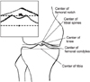
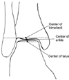
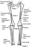
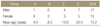
We enrolled 32 patients who were diagnosed as having hereditary multiple exostoses (HME) between January 2001 and December 2007. Based on age at diagnosis, we categorized them into 4 groups, A (0-5 years: 6 patients), B (6-10 years: 7 patients), C (11-15 years: 7 patients) and D (>16 years: 12 patients). We measured mechanical axis deviation, This included femorotibial mechanical angle (a), inferolateral angle (b), femoral mechanical proximal anatomical angle (c), femoral mechanical distal anatomical angle (d), distal tibia inferolateral angle (e) and femoral neck-shaft angle (f). We analyzed for differences among the groups of different ages.
Results
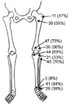
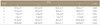
The average femorotibial mechanical angles (a) of Groups A/B/C/D were respectively, 178.5°/180.3°/182.5°/183.5° (p<0.05). Distal tibia inferolateral angles (e) were respectively, 91.9°/93.5°/94.2°/102.9° (p<0.05). The mechanical axis deviation of groups A, B, C, and D, respectively, were 1.7 mm, 6.0 mm, 9.6 mm, and 13.4 mm (p<0.05) on the right side, and 2.9 mm, 7.6 mm, 12.2 mm, and 15.2 mm (p<0.05) on the left side.
Figures and Tables


References
1. Boyer A. Traite des malaidies chirurgicals et des operation qui leur conviennent. 1814. 3. Paris: ve Mignere;594.
2. Harris H. A sex-limiting modifying gene in diaphysial aclasis. Ann Eugen. 1948. 14:165–170.
3. Pierz KA, Stieber JR, Kusumi K, Dormans JP. Hereditary multiple exostoses: one center's experience and review of etiology. Clin Orthop Relat Res. 2002. 401:49–59.

4. Alvarez CM, De Vera MA, Heslip TR, Casey B. Evaluation of the anatomic burden of patients with hereditary multiple exostoses. Clin Orthop Relat Res. 2007. 462:73–79.
5. Solomon L. Hereditary Multiple Exostosis. J Bone Joint Surg Br. 1963. 45:292–304.
6. Hennekam RC. Hereditary multiple exostoses. J Med Genet. 1991. 28:262–266.
7. Nawata K, Teshima R, Minamizaki T, Yamamoto K. Knee deformities in multiple hereditary exostoses. A longitudinal radiographic study. Clin Orthop Relat Res. 1995. 313:194–199.
8. Schmale GA, Conrad EU 3rd, Raskind WH. The natural history of hereditary multiple exostoses. J Bone Joint Surg Am. 1994. 76:986–992.
9. Black B, Dooley J, Pyper A, Reed M. Multiple hereditary exostoses. An epidemiologic study of an isolated community in Manitoba. Clin Orthop Relat Res. 1993. 287:212–217.
10. Krooth RS, Macklin MT, Hilbish TF. Diaphysealaclasis (multiple exostosis) on Guam. Am J Hum Genet. 1961. 13:340–347.
11. McCornack EB. The surgical management of hereditary multiple exostosis. Orthop Rev. 1981. 10:57.
12. Dias LS. Valgus deformity of the ankle joint: Pathogenesis of fi bular shortening. J Pediatr Orthop. 1985. 5:176–180.
13. Jahss MH, Olives R. The foot and ankle in multiple hereditary exostoses. Foot Ankle. 1980. 1:128–142.
14. Shapiro F, Simon S, Glimcher MJ. Hereditary multiple exostoses. Anthropometric, roentgenographic, and clinical aspects. J Bone Joint Surg Am. 1979. 61:815–824.
15. Snearly WN, Peterson HA. Management of ankle deformities in multiple hereditary osteochondromata. J Pediatr Orthop. 1989. 9:427–432.
16. Burgess RC, Cates H. Deformities of the forearm in patients who have multiple cartilaginous exostosis. J Bone Joint Surg Am. 1993. 75:13–18.
17. Fogel GR, McElfresh EC, Peterson HA, Wicklund PT. Management of deformities of the forearm in multiple hereditary osteochondromas. J Bone Joint Surg Am. 1984. 66:670–680.
18. Lee SJ, Joo SD, Park SW, Lee KS, Sung JJ, Lee SH. The appearance and the axial alignment of the lower extremity. J Korean Orthop Assoc. 2004. 39:753–758.
19. Langenskiold A. Normal and pathological bone growth in the light of the development of cartilaginous foci in chondrodysplasia. Acta Chir Scand. 1947. 95:367–381.
20. Siffert RS, Levy RN. Correction of wrist deformity in diaphysealaclasis by stapling. Report of a case. J Bone Joint Surg Am. 1965. 47:378–380.
21. Moreland JR, Bassett LW, Hanker GJ. Radiographic an alysis of the axial alignment of the lower extremity. J Bone Joint Surg Am. 1987. 69:745–749.
22. Jaffe HL. Hereditary multiple exostoses. Arch Pathol. 1943. 36:335–357.
23. Wicklund CL, Pauli RM, Johnston D, Hecht JT. Natural history study of hereditary multiple exostoses. Am J Med Genet. 1995. 55:43–46.
24. Edeikin J. Roentgen diagnosis of diseases of bone. 1981. 3rd Ed. Baltimore: Williams and Wilkins;1402–1409.
25. Kim ID, Lee SY, Ihin JC, Park BC, Ban JY. A case of hereditary multiple osteochondromatosis. J Korean Orthop Assoc. 1982. 17:1005–1010.
26. Turek SL. Orthopaedics, principles and their applications. 1977. 3rd Ed. Philadelphia: J. B. Lippincott Co.;321–322.
27. Rigal WM. Tritiated thymidine in studies of chondrogenesis. J Bone Joint Surg Br. 1961. 43:180.
28. Noonan KJ, Feinberg JR, Levenda A, Snead J, Wurtz LD. Natural history of multiple hereditary osteochondromatosis of the lower extremity and ankle. J Pediatr Orthop. 2002. 22:120–124.
29. Porter DE, Benson MK, Hosney GA. The hip in hereditary multiple exostoses. J Bone Joint Surg Br. 2001. 83:988–995.
 XML Download