

 PDF
PDF ePub
ePub Citation
 Print
Print


Abstract
Purpose
Planovalgus deformity in children with generalized ligamentous laxity is usually asymptomatic, but it sometimes causes severe deformity and functional problems. The aim of this study was to evaluate, post-operatively, functional outcomes, plantar pressure and radiographic results of symptomatic planovalgus with generalized ligamentous laxity.
Materials and Methods
This retrospective study included a total of 42 feet of 24 patients that had undergone a calcaneal lengthening osteotomy or an extra-articular subtalar arthrodesis. The mean age of the patients at the time of the index operation was 10.5 years (range, 6-15.6 years), and the mean duration of follow-up was 51 months (range, 18-92 months). Patients were evaluated clinically and radiographically using the Oxford Ankle Foot Questionnaire, AOFAS score, Mosca criteria, standard radiographs and dynamic pedobarographs.
Results
Functional outcomes at the latest available follow-up were excellent except for three feet. No foot had a significant limitation affecting the patient's daily activities. Both surgical treatments improved radiographic parameters. The parameters of dynamic pedobarographs, including the relative vertical impulse and the peak pressure, decreased for the medial aspect of the forefoot and midfoot, while they increased for the lateral aspect of the forefoot, midfoot and calcaneus after surgical treatment. The change in the center of pressure indicated a significant lateral shift in the weight-bearing surface of the foot.
Conclusion
Calcaneal lengthening osteotomy and extra-articular subtalar arthrodesis appear to be effective means for pain relief and clinical improvement in children with symptomatic planovalgus deformity and generalized ligamentous laxity. Further follow-up evaluation is needed to obtain long-term clinical and radiographic results with regard to skeletal maturation and changes in generalized ligamentous laxity.
Figures and Tables
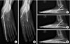 | Figure 1(A) Anteroposterior talo-1st metatarsal angle. (B) Anteroposterior talocalcaneal angle. (C) Lateral talo-1st metatarsal angle. (D) Lateral talocalcaneal angle. (E) Calcaneal pitch.
|
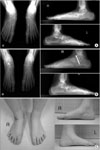 | Figure 2(A) A ten-year-old girl underwent calcaneal lengthening osteotomy 5 years ago. Although radiographs of the left foot show acceptable findings, those of the right foot show loss of reduction and increased anteroposterior and lateral talo-1st metatarsal angle. (B) These radiographs show the decreased anteroposterior and lateral talo-1st metatarsal angle and improved alignment after extra-articular subtalar arthrodesis for the right foot. (C) Although the clinical scores were not excellent, clinical photographs of the patient show acceptable appearances.
|


References
1. Mosca VS. Flexible flatfoot and skewfoot. Instr Course Lect. 1996. 45:347–354.

2. Cappello T, Song KM. Determining treatment of flatfeet in children. Curr Opin Pediatr. 1998. 10:77–81.
3. Tudor A, Ruzic L, Sestan B, Sirola L, Prpic T. Flat-footedness is not a disadvantage for athletic performance in children aged 11 to 15 years. Pediatrics. 2009. 123:e386–e392.
4. Lindsey JM, Michelson JD, MacWilliams BA, Sponseller PD, Miller NH. The foot in Marfan syndrome: clinical findings and weight-distribution patterns. J Pediatr Orthop. 1998. 18:755–759.
5. Wynne-Davies R. Familial joint laxity. Proc R Soc Med. 1971. 64:689–690.
6. Bourelle S, Cottalorda J, Gautheron V, Chavrier Y. Extra-articular subtalar arthrodesis. A long-term follow-up in patients with cerebral palsy. J Bone Joint Surg Br. 2004. 86:737–742.
7. Dennyson WG, Fulford GE. Subtalar arthrodesis by cancellous grafts and metallic internal fixation. J Bone Joint Surg Br. 1976. 58-B:507–510.
8. Evans D. Calcaneo-valgus deformity. J Bone Joint Surg Br. 1975. 57:270–278.
9. Grice DS. An extra-articular arthrodesis of the subastragalar joint for correction of paralytic flat feet in children. J Bone Joint Surg Am. 1952. 34 A:927–940.
10. Mosca VS. Calcaneal lengthening for valgus deformity of the hindfoot. Results in children who had severe, symptomatic flatfoot and skewfoot. J Bone Joint Surg Am. 1995. 77:500–512.
11. Harris RI, Beath T. Hypermobile flat-foot with short tendo achillis. J Bone Joint Surg Am. 1948. 30A:116–140.
12. Wenger DR, Mauldin D, Speck G, Morgan D, Lieber RL. Corrective shoes and inserts as treatment for flexible flatfoot in infants and children. J Bone Joint Surg Am. 1989. 71:800–810.
13. Koutsogiannis E. Treatment of mobile flat foot by displacement osteotomy of the calcaneus. J Bone Joint Surg Br. 1971. 53:96–100.
14. Cohen-Sobel E, Giorgini R, Velez Z. Combined technique for surgical correction of pediatric severe flexible flatfoot. J Foot Ankle Surg. 1995. 34:183–194.
15. Ilyas I, Wade WJ, Al Barrag M, Al Hussainan TS, Lotaibi LA, Alssayad M. A rare pentad of foot and ankle deformities in hyperlax children. J Child Orthop. 2009. 3:115–120.
16. Beighton P, Solomon L, Soskolne CL. Articular mobility in an African population. Ann Rheum Dis. 1973. 32:413–418.
17. Kitaoka HB, Alexander IJ, Adelaar RS, Nunley JA, Myerson MS, Sanders M. Clinical rating systems for the ankle-hindfoot, midfoot, hallux, and lesser toes. Foot Ankle Int. 1994. 15:349–353.
18. Morris C, Doll HA, Wainwright A, Theologis T, Fitzpatrick R. The Oxford ankle foot questionnaire for children: scaling, reliability and validity. J Bone Joint Surg Br. 2008. 90:1451–1456.
19. Davids JR, Gibson TW, Pugh LI. Quantitative segmental analysis of weight-bearing radiographs of the foot and ankle for children: normal alignment. J Pediatr Orthop. 2005. 25:769–776.
20. Vanderwilde R, Staheli LT, Chew DE, Malagon V. Measurements on radiographs of the foot in normal infants and children. J Bone Joint Surg Am. 1988. 70:407–415.
21. Kirtley C. Kirtley C, editor. Plantar pressure measurement. 2006. Oxford: Churchill Livingstone;97–115.
22. Grahame R. The hypermobility syndrome. Ann Rheum Dis. 1990. 49:199–200.
23. McCall RE, Lillich JS, Harris JR, Johnston FA. The Grice extraarticular subtalar arthrodesis: a clinical review. J Pediatr Orthop. 1985. 5:442–445.
24. Cooper PS, Nowak MD, Shaer J. Calcaneocuboid joint pressures with lateral column lengthening (Evans) procedure. Foot Ankle Int. 1997. 18:199–205.
25. Dinucci KR, Christensen JC, Dinucci KA. Biomechanical consequences of lateral column lengthening of the calcaneus: Part I. Long plantar ligament strain. J Foot Ankle Surg. 2004. 43:10–15.
26. André M, Hagelberg S, Stenström CH. The juvenile arthritis foot disability index: development and evaluation of measurement properties. J Rheumatol. 2004. 31:2488–2493.
27. Roye BD, Vitale MG, Gelijns AC, Roye DP Jr. Patient-based outcomes after clubfoot surgery. J Pediatr Orthop. 2001. 21:42–49.
28. Park KB, Park HW, Lee KS, Joo SY, Kim HW. Changes in dynamic foot pressure after surgical treatment of valgus deformity of the hindfoot in cerebral palsy. J Bone Joint Surg Am. 2008. 90:1712–1721.
29. Ledoux WR, Hillstrom HJ. The distributed plantar vertical force of neutrally aligned and pes planus feet. Gait Posture. 2002. 15:1–9.
 XML Download