

 PDF
PDF ePub
ePub Citation
 Print
Print


Abstract
Purpose
We wanted to analyze the results of the 1st metatarsal dorsal close wedge osteotomy (MTDW) combined with medical cuneiform plantar open wedge (MCPOW) for treating forefoot deformity of a cavus foot.
Materials and Methods
We retrospectively analyzed 30 patients. Their mean age was 21.5 years (SD 10.6 years) and the average follow-up period was 2.3 years. Thirty-four cases of thirty patients were classified as group A, as classified by the 1st MTDW combined with the MCPOW, 16 feet (14 patients) were group B by the 1st MTDW or MCPOW, 12 feet (10 patients), and group C by triple arthrodesis, 6 feet (6 patients). We evaluated the ankle dorsiflexion, plantarflexion, heel alignment, and the Maryland foot score (MFS) preoperatively and the last follow-up, and we analyzed the radiologic Hibb, Meary, calcaneal pitch and tibiotalar angles.
Results
The ankle dorsiflexion (p=0.01), plantar flexion (p=0.03) and heel alignment (p=0.02) of group A were significantly improved more than that of groups B and C. The MFS of group A revealed better than group B and C (p=0.01). The Meary (p=0.01), Hibb (p=0.02) and calcaneal pitch angle (p=0.02) of group A were significantly improved more than that of groups B and C.
Figures and Tables
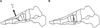 | Figure 1Schematic drawing illustrating demonstrated the double osteotomy of forefoot. (A) Dorsal wedge osteotomy of the first metatarsal bone followed by obtaining a wedge shaped segmental bone. (B) Plantar osteotomy of the medial cuneiform followed by insertion a bone fragment.
|
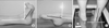 | Figure 2Clinical photographs of patient with cavus deformity. Measurement of ankle angle on dorsiflexion, (A) and plantarflexion, (B) and heal alignment angle between hindfoot and ground, (C).
|
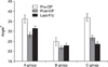
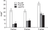 | Figure 3Histogram demonstrate mean Meary's angle at preoperative and follow-up period in three groups, and the angle of group A was best corrected than other groups (p=0.01).
|
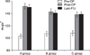 | Figure 4Histogram demonstrate mean Hibb's angle at preoperative and follow-up period in three groups, and the angle of group A was best corrected than other groups (p=0.02).
|
 | Figure 5Histogram demonstrate mean calcaneal pitch angle at preoperative and follow-up period in three groups, the angle of group A was best corrected than other groups (p=0.02).
|
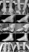 | Figure 6(A) Clinical photographs of male patient with Charcot-Marie-Tooth disease. (B) Preoperative lateral radiograph demonstrating abnormal Meary's, Hibb's and calcaneal pitch angle. (C) Operative photograph showing double osteotomy of forefoot, and grafting segmental bone to plantar osteotomy site of the medial cuneiform. (D) Lateral radiograph made 2 years after osteotomy demonstrating correction of cavus deformity. (E) Photograph made 2 years after osteotomy showing that the ankle dorsiflexion, plantarflexion and healignment angle was improved than preoperative period.
|
References
1. Mosca VS. The cavus foot. J Pediatr Ortho. 2001. 21:423–424.

2. Paulos L, Coleman SS, Samuelson KM. Pes cavovarus. Review of a surgical approach using selective soft-tissue procedures. J Bone Joint Surg Am. 1980. 62:942–953.
3. Sabir M, Lyttle D. Pathogenesis of pes cavus in Charcot-Marie-Tooth disease. Clin Orthop Relat Res. 1983. 175:173–178.
4. Alexander IJ, Johnson KA. Assessment and management of pes cavus in Charcot-Marie-tooth disease. Clin Orthop Relat Res. 1989. 246:273–281.
5. Azmaipairashvili Z, Riddle EC, Scavina M, Kumar SJ. Correction of cavovarus foot deformity in Charcot-Marie-Tooth disease. J Pediatr Orthop. 2005. 25:360–365.
6. Sanders R, Fortin P, DiPasquale T, Walling A. Operative treatment in 120 displaced intra articular calcaneal fractures. Results using a prognostic computed tomography scan classification. Clin Orthop Relat Res. 1993. 290:87–95.
7. Sherman FC, Westin GW. Plantar release in the correction of deformities of the foot in childhood. J Bone Joint Surg Am. 1981. 63:1382–1389.
8. Jahss MH. Tarsometatarsal truncated-wedge arthrodesis for pes cavus and equinovarus deformity of the fore part of the foot. J Bone Joint Surg Am. 1980. 62:713–722.
9. Wicart P, Seringe R. Plantar opening-wedge osteotomy of cuneiform bones combined with selective plantar release and dwyer osteotomy for pes cavovarus in children. J Pediatr Orthop. 2006. 26:100–108.
10. Medhat MA, Karantz H. Neuropathic ankle joint in Charcot-Marie-Tooth disease after triple arthrodesis of the foot. Ortho Rev. 1988. 17:873–880.
11. Wukich DK, Bowen JR. A long-term study of triple arthrodesis for correction of pes cavovarus in Charcot-Marie-Tooth disease. J Pediatr Orthop. 1989. 9:433–437.
12. Mubarak SJ, Van Valin SE. Osteotomies of the foot for cavus deformities in children. J Pediatr Orthop. 2009. 29:294–299.
13. Coleman SS, Chestnut WJ. A Simple test for hindfoot flexibility in the cavovarus foot. Clin Orthop. 1977. 123:60–62.
14. Dwyer FC. Osteotomy of the calcaneum for pes cavus. J Bone Joint Surg Br. 1959. 41:80–86.
15. Jahss MH. Evaluation of the cavus foot for orthopedic treatment. Clin Orthop Relat Res. 1983. 181:52–63.
16. Schwend RM, Drennan JC. Cavus foot deformity in children. J Am Acad Orthop Surg. 2003. 11:201–211.
17. Giannini S, Ceccarelli F, Benedetti MG, Faldini C, Grandi G. Surgical treatment of adult idiopathic cavus foot with plantar fasciotomy, naviculocuneiform arthrodesis, and cuboid osteotomy. A review of thirty-nine cases. J Bone Joint Surg Am. 2002. 84:Suppl. 62–69.
18. Mann DC, Hsu JD. Triple arthrodesis in the treatment of fixed cavovarus deformity in adolescent patients with Charcot-Marie-Tooth disease. Foot Ankle. 1992. 13:1–6.
19. Wetmore RS, Drennan JC. Long-term results of triple arthrodesis in Charcot-Marie-Tooth disease. J Bone Joint Surg Am. 1989. 71:417–422.
20. Gould N. Surgery in advanced Charcot-Marie-Tooth disease. Foot Ankle. 1984. 4:267–273.
21. Japas LM. Surgical treatment of pes cavus by tarsal V-osteo-tomy. Preliminary report. J Bone Joint Surg Am. 1968. 50:927–944.
 XML Download