

 PDF
PDF ePub
ePub Citation
 Print
Print


Abstract
Purpose
Recently, the methods for reconstructing the acromion joint focused on an anatomical reconstruction. However, the reports of the anatomical method of the coraco-clavicle ligament have given different descriptions. This paper reports an anatomical study of the coraco-clavicle ligament in Koreans.
Materials and Methods
One hundred and two coraco-clavicle ligaments distracted from 6 fresh cadavers and 96 drug-administered cadavers were analyzed and calibrated. A quantitative analysis of the shape and location of the attachment site of the coracoid process and clavicle was performed, and the clinical and radiological applications were examined considering the calibrating variables.
Results
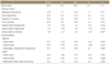
The conoid and trapezoid ligaments were thick and wide when measured in the middle part, and the average length up to the attachment site of the coracoid process was 11.3±3.6mm in men and 9.5±2.2mm in women. The average length of the clavicle was 158.5±55.7mm in men and 146.6±50.9mm in women. The length between the middle of the trapezoid tubercle or conoid tubercle and the lateral edge of the clavicle was 21.7±3.2mm and 42.8±3.4mm respectfully in men and 20.9±1.8mm and 39.9±3.3 respectfully in women, respectively. The angles of the conoid ligament and trapezoid ligament from the anteroposterior and lateral aspects were measured to be 25±8° and 19±3° respectfully in men and 28±5° and 17±3° degree respectfully in women.
Figures and Tables
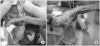
Figure 1
(A) The distance from the lateral end of the clavicle to the lateral extent of the trapezoid ligament (a) and to the medial end of the trapezoid ligament (b). (B) The distance from the lateral end of the clavicle to the lateral extent of the conoid ligament (c) and to the medial end of the conoid ligament (d).
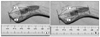
Figure 2
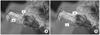
The distance from the posterior cortex of the clavicle to (a) the posterior extent of the trapezoid ligament (b) the anterior extent of the trapezoid ligament (c) the posterior extent of the conoid ligament (d) the anterior extent of the conoid ligament.
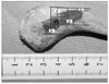
Figure 3
(A) The width (a) and the thickness (b) measured at the midsubstance of the trapezoid ligament. (B) The width (a) and the thickness (b) measured at the midsubstance of the conoid ligament.
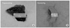
Figure 4
(A) The distance from the tip of coracoid to the anterior extent of the trapezoid ligament (a) and to the posterior extent of the trapezoid ligament (b). (B) The distance from the tip of coracoid to the anterior extent of the conoid ligament (c) and to the posterior extent of the conoid ligament (d).


References
1. Fukuda K, Craig EV, An KN, Cofield RH, Chao EY. Biomechanical study of the ligamentous system of the acromioclavicular joint. J Bone Joint Surg Am. 1986. 68:434–440.

2. Lee KW, Debski RE, Chen CH, Woo SL, Fu FH. Functional evaluation of the ligaments at the acromioclavicular joint during anteroposterior and superoinferior translation. Am J Sports Med. 1997. 25:858–862.
3. Costic RS, Labriola JE, Rodosky MW, Debski RE. Biomechanical rationale for development of anatomical reconstructions of coracoclavicular ligaments after complete acromioclavicular joint dislocations. Am J Sports Med. 2004. 32:1929–1936.
4. Salter EG Jr, Nasca RJ, Shelley BS. Anatomical observations on the acromioclavicular joint and supporting ligaments. Am J Sports Med. 1987. 15:199–206.
5. Bannister GC, Wallace WA, Stableforth PG, Hutson MA. The management of acute acromioclavicular dislocation. A randomised prospective controlled trial. J Bone Joint Surg Br. 1989. 71:848–850.
6. Rios CG, Arciero RA, Mazzocca AD. Anatomy of the clavicle and coracoid process for reconstruction of the coracoclavicular ligaments. Am J Sports Med. 2007. 35:811–817.
7. Rockwood CA Jr, Williams GR, Young DC. Rockwood CA, Malsen FA, editors. Disorders of the acromioclavicular joint. 1998. 2nd ed. Philadelphia, Pa: WB Sanders Co;483–553.
8. Kent BE. Functional anatomy of the shoulder complex. A review. Phys Ther. 1971. 51:947.
9. Harris RI, Wallace AL, Harper GD, Goldberg JA, Sonnabend DH, Walsh WR. Structural properties of the intact and the reconstructed coracoclavicular ligament complex. Am J Sports Med. 2000. 28:103–108.
10. Motamedi AR, Blevins FT, Willis MC, McNally TP, Shahinpoor M. Biomechanics of the coracoclavicular ligament complex and augmentations used in its repair and reconstruction. Am J Sports Med. 2000. 28:380–384.
11. Grutter PW, Petersen SA. Anatomical acromioclavicular ligament reconstruction: a biomechanical comparison of reconstructive techniques of the acromioclavicular joint. Am J Sports Med. 2005. 33:1723–1728.
12. Mazzocca AD, Conway JE, Johnson S, et al. The anatomic coracoclavicular ligament reconstruction. Oper Tech Sports Med. 2004. 12:56–61.
13. Flatow EL, Cordasco FA, Bigliani LU. Arthroscopic resection of the outer end of the clavicle from a superior approach: a critical, quantitative, radiographic assessment of bone removal. Arthroscopy. 1992. 8:55–64.
14. Blazar PE, Iannotti JP, Williams GR. Anteroposterior instability of the distal clavicle after distal clavicle resection. Clin Orthop Relat Res. 1998. 348:114–120.
 XML Download