

 PDF
PDF ePub
ePub Citation
 Print
Print


Abstract
Purpose
Recently, there have been many attempts to use PET-CT for the diagnosis of metastasis of soft tissue tumors and for differentiating benign tumors from malignant tumors. In this study, we wanted to evaluate the efficacy of 18-FDG PET CT in soft tissue tumors.
Materials and Methods
Patients (n=64) with suspected malignant soft tissue tumor had 18-FDG PET CT scans. This included 15 cases of benign soft tissue tumor, 34 cases of malignant soft tissue tumor and 15 cases of inflammatory lesions. All patients went through surgical treatment after PET-CT imaging was done, and all dissected tissues were biopsied. For semiquantitive analysis, SUVmax(Maximal standard uptake value) was measured, and SUVmax was determined using ROC analysis, the Kruskal-Wallis test and the Mann-Whitney test.
Results
Using 18-FDG PET CT it was possible to differentiate benign from malignant tumor using SUVmax values. But, discrimination between different grades of tumor was not possible. Also discrimination between malignant tumors and inflammatory lesions was not possible.
Conclusion
PET-CT can be considered as a useful nuclear imaging method that can detect local recurrence and distant metastasis of soft tissue sarcoma and can differentiate benign from malignant tumors. But PET-CT results must be interpreted carefully in diagnoses when there is inflammatory disease or a suspected lesion present.
Figures and Tables
Figure 1
Statistical data for patient subset based on tumor grade (Benign lesion, Grade I, Grade II, Grade III, Inflammatory lesion).
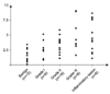
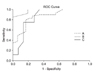
Figure 2
Comparison of ROC curves for differeniation with soft tissue sarcoma and benign soft tissue mass (AUC, 0.872; SE, 0.057; 95% CI, 0.760-0.983; p=0.000) (A), differentiation with liposarcoma and lipoma (AUC, 0977; SE, 0.031; 95% CI, 0.916-1.038; p=0.002) (B), and differentiation with low grade soft tissue sarcoma and benign soft tissue mass (AUC, 0858; SE, 0.079; 95% CI, 0.704-1.012; p=0.006) (C).

References
1. Iagaru A, Quon A, McDougall IR, Gambhir SS. F-18 FDG PET/CT evaluation of osseous and soft tissue sarcomas. Clin Nucl Med. 2006. 31:754–760.

2. Dimitrakopoulou-Strauss A, Strauss LG, Schwarzbach M, et al. Dynamic PET 18F-FDG studies in patients with primary and recurrent soft -tissue sarcomas: impact on diagnosis and correlation with grading. J Nucl Med. 2001. 42:713–720.
3. Coindre JM. Grading of soft tissue sarcomas: review and update. Arch Pathol Lab Med. 2006. 130:1448–1453.
4. Ernest U, Conrad M, Hannah D, et al. Fluorodeoxyglucose positron emission tomography scanning: basic principles and imaging of adult soft-tissue sarcomas. J Bone Joint Surg Am. 2004. 86:98–104.
5. Schöder H, Erdi YE, Chao K, Gonen M, Larson SM, Yeung HW. Clinical implications of different image reconstruction parameters for interpretation of whole-body PET studies in cancer patients. J Nucl Med. 2004. 45:559–566.
6. Aoki J, Watanabe H, Shinozaki T, et al. FDG-PET for preoperative differential diagnosis between benign and malignant soft tissue masses. Skeletal Radiol. 2003. 32:133–138.
7. Lucas JD, O'Doherty MJ, Wong JC, et al. Evaluation of fluorodeoxy glucose positron emission tomography in the management of soft-tissue sarcomas. J Bone Joint Surg Br. 1998. 80:441–447.
8. Hamada K, Tomita Y, Ueda T, et al. Evaluation of delayed 18F-FDG PET in differential diagnosis for malignant soft-tissue tumors. Ann Nucl Med. 2006. 20:671–675.
9. Schwarzbach MH, Dimitrakopoulou-Strauss A, Willeke F, et al. Clinical value of [18-F] fluorodeoxyglucose positron emission tomography imaging in soft tissue sarcomas. Ann Surg. 2000. 231:380–386.
10. Nieweg OE, Pruim J, van Ginkel RJ, et al. Fluorine-18-fluorodeoxyglucose PET imaging of soft-tissue sarcoma. J Nuc Med. 1996. 37:257–261.
11. Salanova V, Markland O, Woth R. Longitudinal follow-up in 145 patients with medically refractory temporal lobe epilepsy treated surgically between 1984 and 1995. Epilepsia. 1999. 40:1417–1423.
12. Smith MA, O'Doherty MJ. Position emission tomography and the orthopaedic surgeon. J Bone Joint Surg Br. 2004. 82:324–325.
13. Feldman F, van Heertum R, Manos C. 18 FDG PET scanning of benign and malignant musculoskeletal lesions. Skeletal Radiol. 2003. 32:201–208.
14. Tateishi U, Yamaguchi U, Seki K, Terauchi T, Arai Y, Kim EE. Bone and soft-tissue sarcoma: preoperative staging with fluorine 18 fluorodeoxyglucose PET/CT and conventional imaging. Radiology. 2007. 245:839–847.
 XML Download