

 PDF
PDF ePub
ePub Citation
 Print
Print


Abstract
Purpose
To report the loss of correction of a sagittal imbalance and the clinical outcomes after a corrective osteotomy for lumbar degenerative kyphosis.
Materials and Methods
This study analyzed the radiological parameters, surgical techniques, and clinical outcomes of 23 patients, who underwent corrective osteotomy for lumbar degenerative kyphosis. The patients were divided into groups I (>5 cm loss of correction of sagittal imblance, 12 patients) and II (<5 cm, 11 patients) to compare the patients with the correction preserved with those with the correction lost. In terms of the clinical outcome, group A (high satisfaction score group >3.5 out of 5, 11 patients) was compared with group B (low satisfaction score group <3.5 out of 5, 12 patients).
Results
The sagittal imbalance was corrected by performing a Smith-Petersen osteotomy (SPO) in 11 cases and Pedicle subtraction osteotomy (PSO) in 12. The mean preoperative sagittal imbalance was improved from 26.4 cm to 4.05 cm, postoperatively, and 11.2 cm at the last follow up. The mean loss of correction was 11.2 cm in group I and 2.3 cm in group II. The mean satisfaction score was 4.56 in group A and 2.18 in group B. The presence of an old compression fracture was found to be related to the loss of correction, and the preoperative symptomatic spinal stenosis was related to poor clinical outcomes.
Figures and Tables
Fig. 1
Radiographs after the Smith-Peterson osteotomy with anterior lumbar interbody fusion (A) and Pedicle subtraction osteotomy (B) in lumbar degenerative kyphosis patients.
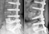
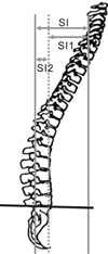
Fig. 3
Radiographs showing LDK patients with reactive thoracic lordosis (A) and without reactive thoracic lordosis (B).
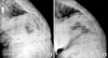
Fig. 4
Serial radiographs of a 72-year-old female patient showing loss of sagittal balance. (A) Preoperative radiograph showing severe sagittal imbalance (35.5 cm). (B) Immediate postoperative radiographs showing a restoration of the sagittal balance (4.5 cm). Note the line from the posterosuperior corner of S1 to the center of the T12/L1 disc passes in front of T1. (C) Two years after surgery, the sagittal balance was lost (19.2 cm). The same line of (B) passes in back of T1. This suggests that a significant loss of correction occurred at the proximal unfused segments.
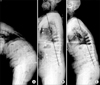
Fig. 5
Serial radiographs of a 74-year-old female patient showing severe loss of correction 46 months after surgery. Most of the loss occurred at proximal unfused segment with degenerative changes.
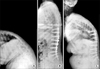
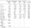
Table 2
Analysis of the Related Factors in the Patients Showing a Preserved (Group I) and Loss (Group II) of Sagittal Balance
*SI, sagittal imbalance; †PI, pelvic incidence; ‡SS, sacral slope; §PVM, paravertebral muscle cross-section area; ∥OCF, old compression fracture; ¶BDM, Mean lumbar spine bone densitometry; **SPO, Smith-Peterson Osteotomy; PSO, Pedicle subtraction osteotomy; **BDM, Mean lumbar spine bone densitometry; ††MSRSI, Modified Spine Research Society Instrument; ‡‡RTL, Reactive thoracic lordosis.
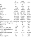
Table 3
Analysis of the Related Factors in the Patients Showing Good (Group A) and Poor (Group B) Satisfaction
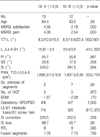
*MSRSI, Modified Spine Research Society Instrument; †PI, Pelvic Incidence; ‡SS, Sacral Slope; §SI, Sagittal Index; ∥PVM, Paravertebral Muscle Cross-section Area; ¶OCF, Old Compression Fracture; **BDM, Mean Lumbar Spine Bone Densitometry; ††SPO, Smith-peterson Osteotomy; PSO, Pedicle Subtraction Osteotomy; ‡‡RTL, Reactive Thoracic Lordosis.
References
1. Doherty JH. Complication of fusion in lumbar scoliosis. J Bone Joint Surg Am. 1973. 55:438–449.
2. Jansen RC, Van Rhijn LW, Van Ooij A. Predictable correction of the unfused lumbar lordosis after thoracic correction and fusion in scheuermann kyphosis. Spine. 2006. 31:1227–1231.

3. Kang CH, Shin MJ, Kim SM, Lee SH, Lee CS. MRI of paraspinal muscles in lumbar degenerative kyphosis patients and control patients with chronic low back pain. Clinical Radiology. 2007. 62:479–486.
4. Kim EH, Han SK, Kim HJ. A clinical analysis of surgical treatment of lumbar degenerative kyphosis. J Korean Soc Spine Surg. 2001. 8:210–218.
5. Kim EH, Kim SW. Anterior and posterior surgical treatment with wedged cage (SynCage®) in lumbar degenerative kyphosis. J Korean Spine Surg. 2003. 10:240–247.
6. Kim YJ, Bridwell KH, Lenke LG, Rhim S, Cheh G. Sagittal thoracic decompensation following long adult lumbar spinal instrumentation and fusion to L5 or S1: causes, prevalence, and risk factor analysis. Spine. 2006. 31:2359–2366.
7. Lee CS, Chung SS, Chung KH, Kim SR. Significance of pelvic incidence in the development of abnormal sagittal alignment. J Korean Orthop Assoc. 2006. 41:274–280.
8. Lee CS, Kim YT, Kim E. Clinical study of lumbar degenerative kyphosis. J Korean Spine Surg. 1997. 4:27–35.
9. Lee CS, Lee CK, Kim YT, Hong YM, Yoo JH. Dynamic sagittal imbalance of the spine in degenerative flat back: significance of pelvic tilt in surgical treatment. Spine. 2001. 26:2019–2035.
10. Shufflebarger H, Suk SI, Mardjetko S. Debate: determining the upper instrumented vertebra in the management of adult degenerative scoliosis: stopping at T10 versus L1. Spine. 2006. 31:Suppl 19. S185–S194.
11. Takemitsu Y, Harada Y, Iwahara T, Miyamoto M, Mitatake Y. Lumbar degenerative kyphosis. Clinical, radiological and epidemiological studies. Spine. 1988. 13:1317–1326.
 XML Download