

 PDF
PDF ePub
ePub Citation
 Print
Print


Abstract
Purpose
We evaluated the degree of femoral and tibial torsion in, and the efficacy of two operative procedures for, resistant idiopathic clubfoot with toe-in gait.
Materials and Methods
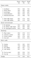
Thirty one feet in 23 patients (average age at the time of revision surgery 4.3 years) were studied. CT was used to determine femoral anteversion and tibial torsion. Two different operative procedures were applied, depending on the degree of toe-in gait: group 1 (10 feet whose toe-in gait was not severe) - soft tissue release, anterior tibial tendon transfer and mid-foot (cuboid closing and cuneiform opening) osteotomy; group 2 (21 feet which had relatively severe toe-in gait) - supramalleolar external rotation osteotomy of the distal tibia (SEROT), along with the same procedure as group 1. Mean follow-up period after revision surgery was 6.3 years. Results were assessed radiologically and clinically with the Dimeglio classification and Clubfoot Assessment Protocol.
Results
The mean femoral anteversion and external-tibial torsion of the affected side were increased. Twenty eight of 31 feet (90.3%) demonstrated excellent or good results. In group 2, we obtained 19 excellent (90.5%) and 2 good (9.5%) results. Group 1 had 6 excellent (60%), one good (10%) and 3 fair (30%) results.
Figures and Tables
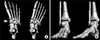 | Fig. 1(A) Three-dimensional illustrations showing midfoot osteotomy. The lateral wedge resection from the cuboid is applied to the medial cuneiform. (B) Improved forefoot adduction and correction of toe-in foot position is seen following the wedge transfer and SEROT.
|
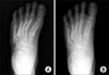 | Fig. 2Weight-bearing AP radiographs of the left clubfoot in a 5-year-old boy who underwent midfoot osteotomy and SEROT: (A) before the secondary surgery (4 years after the initial PMLR) and (B) at the last follow up (3 years after secondary treatment). Reduced forefoot adduction is evident.
|
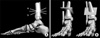 | Fig. 3(A) shows how the osteotomy cut in SEROT may be made incorrectly in the sagittal plane: a cut along line a will result in a varus ankle deformity (B, left side); a cut along line b will result in a valgus ankle deformity (B, right side). The correct cut in Fig. A is indicated by the horizontal line, at right angles to the bone (parallel to the joint line in both the coronal and sagittal planes).
|
References
1. Ponseti IV, Smoley EN. Congenital clubfoot: the results of treatment. J Bone Joint Surg Am. 1963. 45:261–275.
2. Kite JH. Some suggestions on the treatment of clubfoot by casts. J Bone Joint Surg Am. 1963. 45:406–412.
3. Kite JH. The clubfoot. 1964. New York: Grune and Stratton, Inc..
4. Dierauer S, Schafer D, Hefti F. Osteotomies of the mid- and back-foot in recurrent club foot. Orthopade. 1999. 28:117–124.
5. Evans D. Relapsed club foot. J Bone Joint Surg Br. 1961. 43:722–733.

6. Ezra E, Hayek S, Gilai AN, Khermosh O, Wientroub S. Tibialis anterior tendon transfer for residual dynamic supination deformity in treated club feet. J Pediatr Orthop B. 2000. 9:207–211.
7. Garceau GJ. Anterior tibial tendon transposition in recurrent congenital clubfoot. J Bone Joint Surg Am. 1940. 22:932–936.
8. Gupta AK, Kumar R. Treatment of residual club-foot deformity, the bean-shaped foot--by open wedge medial cuneiform osteotomy and closing wedge cuboid osteotomy, clinical review and cadaver correlations. J Pediatr Orthop. 1993. 13:408–410.
9. Hofmann AA, Constine RM, McBride GG, Coleman SS. Osteotomy of the first cuneiform as treatment of residual adduction of the fore part of the foot in club foot. J Bone Joint Surg Am. 1984. 66:985–990.
10. Köse N, Günal I, Göktürk E, Seber S. Treatment of severe residual clubfoot deformity by trans-midtarsal osteotomy. J Pediatr Orthop B. 1999. 8:251–256.
11. Kuo KN, Hennigan SP, Hastings ME. Anterior tibial tendon transfer in residual dynamic clubfoot deformity. J Pediatr Orthop. 2001. 21:35–41.
12. Lovell WW, Winter RB. Pediatric orthopaedics. 2006. Vol. 2:6th ed. Philadelphia: JB Lippincott Company;1262–1277.
13. Pohl M, Nicol RO. Transcuneiform and opening wedge medial cuneiform osteotomy with closing wedge cuboid osteotomy in relapsed clubfoot. J Pediatr Orthop. 2003. 23:70–73.
14. McHale KA, Lenhart MK. Treatment of residual clubfoot deformity--the "bean-shaped" foot--by opening wedge medial cuneiform osteotomy and closing wedge cuboid osteotomy. Clinical review and cadaver correlations. J Pediatr Orthop. 1991. 11:374–381.
15. Farsetti P, Caterini R, Mancini F, Potenza V, Ippolito E. Anterior tibial tendon transfer in relapsing congenital clubfoot: long term follow up study of two series treated with a different protocol. J Pediatr Orthop. 2006. 26:83–90.
16. Diméglio A, Bensahel H, Souchet P, Mazeau P, Bonnet F. Classification of clubfoot. J Pediatr Orthop B. 1995. 4:129–136.
17. Andriesse H, Roos EM, Hägglund G, Jarnol GB. Validity and responsiveness of the clubfoot assessment protocol (CAP). A methodological study. BMC Musculoskelet disord. 2006. 7:28.
18. Cummings RJ, Davidson RS, Armstrong PF, Lehman WB. Congenital clubfoot. J Bone Joint Surg Am. 2002. 84:290–308.
19. Kim HT, Cho JY, Cheon SJ, Yoo CI. The outcomes of surgical treatment for idiopathic clubfoot. J Korean Orthop Assoc. 2005. 40:970–976.
20. Herzenberg JE, Radler C, Bor N. Ponseti versus traditional methods of casting for idiopathic clubfoot. J Pediatr Orthop. 2002. 22:517–521.
21. Morcuende JA, Dolan LA, Dietz FR, Ponseti IV. Radical reduction in the rate of extensive corrective surgery for clubfoot using the ponseti method. Pediatrics. 2004. 113:376–380.
22. Cummings RJ, Lovell WW. Operative treatment of congenital idiopathic club foot. J Bone Joint Surg Am. 1988. 70:1108–1112.
23. Simons GW. Analytical radiography of club feet. J Bone Joint Surg Br. 1977. 59:485–489.
24. el-Tayeby HM. The neglected clubfoot: a salvage procedure. J Foot Ankle Surg. 1998. 37:501–509.
25. Mckay DW. New concept of and approach to clubfoot treatment: section I-principles and morbid anatomy. J Pediatr Orthop. 1982. 2:347–356.
26. Krishina M, Evans R, Sprigg A, Taylor JF, Thesis JC. Tibial torsion measured by ultrasound in children with talipes equinovarus. J Bone Joint Surg Br. 1991. 73:207–210.
27. Reikerås O, Kristiansen LP, Gunderson R, Steen H. Reduced tibial torsion in congenital clubfoot. CT measurements in 24 patients. Acta Orthop Scand. 2001. 72:53–56.
28. Cuevas de Alba C, Guille JT, Bowen JR, Harcke HT. Computed tomography for femoral and tibial torsion in children with clubfoot. Clin Orthop Relat Res. 1998. 353:203–209.
29. Lloyd-Roberts GC, Swann M, Catterall A. Medial rotational osteotomy for severe residual deformity in club foot. A preliminary report on a new method of treatment. J Bone Joint Surg Br. 1974. 56:37–44.
30. Lichtblau S. External rotation tibial osteotomy in clubfoot: adverse late effect. Clin Orthop Relat Res. 1978. 136:225–229.
 XML Download