

 PDF
PDF ePub
ePub Citation
 Print
Print


Abstract
Purpose
This study examined the value and indications of repeated MRI in degenerative lumbar diseases under conservative management by comparing the primary MR and repeated MR images with respect to the symptomatic and radiological changes.
Materials and Methods
Seventy patients with degenerative lumbar disease under conservative management underwent repeat MRI. Five MRI findings, including disc, foramen, facet joint, nerve root, and ligamentum flavum, were used to examine the difference between the initial and repeat MRI. The severity was graded using a four-point scale for each item. The patients were divided into 3 groups in order to compare the radiological changes and symptomatic changes, as follows; Group I no change from the initial symptoms (38 cases), Group II aggravation of the initial pain (18 cases), and Group III aggravation of the initial neurology or the development of a new neurology (14 cases).
Results
The mean scores of each item the disc, foramen, facet joint, nerve root and ligamentum flavum increased from 1.76, 1.31-1.79, 1.71, and 1.47, respectively, to 1.90, 1.47, 1.80, 1.79, and 1.53, respectively. Group III showed the greatest proportion of cases with an increasing grading score (78%, 11 cases) only the disc was significant.
Figures and Tables
Fig. 1
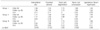
The seventy-three year old male performed repeat MRI on 7 months interval due to Rt. sciatic symptom and motor weakness aggravation (Group III). The grading scores of each parameter were increased from disk 2, foramen 2, facet joint 2, and nerve root 2, ligamentum flavum 1 (A, B) to 3, 3, 2, 3, 1 (C, D) respectively.
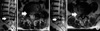
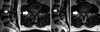
Fig. 2
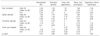
The fifty-nine year old male performed repeat MRI on 14 months interval due to the symptom persistent (Group I). The grading scores of each parameter were decreased from disk 3, foramen 1, facet joint 1, and nerve root 3, ligamentum flavum 1 (A, B) to 2, 1, 1, 2, 1 (C, D) respectively.
References
1. Kirkaldy-Willis WH, Wedge JH, Yong-Hing K, Reilly J. Pathology and pathogenesis of lumbar spondylosis and stenosis. Spine (Phila Pa 1976). 1978. 3:319–328.

2. Yong-Hing K, Kirkaldy-Willis WH. The pathophysiology of degenerative disease of the lumbar spine. Orthop Clin North Am. 1983. 14:491–504.
3. Modic MT, Ross JS. Lumbar degenerative disk disease. Radiology. 2007. 245:43–61.
4. Benoist M. The natural history of lumbar degenerative spinal stenosis. Joint Bone Spine. 2002. 69:450–457.
5. Simotas AC, Dorey FJ, Hansraj KK, Cammisa F Jr. Nonoperative treatment for lumbar spinal stenosis. Clinical and outcome results and a 3-year survivorship analysis. Spine (Phila Pa 1976). 2000. 25:197–203.
6. Saal JA, Saal JS, Herzog RJ. The natural history of lumbar intervertebral disc extrusions treated nonoperatively. Spine. 1990. 15:683–686.
7. Brant-Zawadzki MN, Jensen MC, Obuchowski N, Ross JS, Modic MT. Interobserver and intraobserver variability in interpretation of lumbar disc abnormalities. A comparison of two nomenclatures. Spine (Phila Pa 1976). 1995. 11:1257–1263.
8. Masaryk TJ, Ross JS, Modic MT, Boumphrey F, Bohlman H, Wilber G. High-resolution MR imaging of sequestered lumbar intervertebral disks. AJR Am J Roentgenol. 1988. 5:1155–1162.
9. Wildermuth S, Zanetti M, Duewell S, et al. Lumbar spine: quantitative and qualitative assessment of positional (upright flexion and extension) MR imaging and myelography. Radiology. 1998. 2:391–398.
10. Weishaupt D, Zanetti M, Boos N, Hodler J. MR imaging and CT in osteoarthritis of the lumbar facet joints. Skeletal Radiol. 1999. 4:215–219.
11. Pfirrmann CW, Dora C, Schmid MR, Zanetti M, Hodler J, Boos N. MR image-based grading of lumbar nerve root compromise due to disk herniation: reliability study with surgical correlation. Radiology. 2004. 2:583–588.
12. Sakamaki T, Sairyo K, Sakai T, Tamura T, Okada Y, Mikami H. Measurements of ligamentum flavum thickening at lumbar spine using MRI. Arch Orthop Trauma Surg. 2009. 129:1415–1419.
13. Yukawa Y, Kato F, Matsubara Y, Kajino G, Nakamura S, Nitta H. Serial magnetic resonance imaging follow-up study of lumbar disc herniation conservatively treated for average 30 months: relation between reduction of herniation and degeneration of disc. J Spinal Disord. 1996. 9:251–256.
14. Johnsson KE, Udén A, Rosén I. The effect of decompression on the natural course of spinal stenosis. A comparison of surgically treated and untreated patients. Spine (Phila Pa 1976). 1991. 16:615–619.
15. Simotas AC. Nonoperative treatment for lumbar spinal stenosis. Clin Orthop Relat Res. 2001. 384:153–161.
16. Cribb GL, Jaffray DC, Cassar-Pullicino VN. Observations on the natural history of massive lumbar disc herniation. J Bone Joint Surg Br. 2007. 89:782–784.
17. Awad JN, Moskovich R. Lumbar disc herniations: surgical versus nonsurgical treatment. Clin Orthop Relat Res. 2006. 443:183–197.
18. Bozzao A, Gallucci M, Masciocchi C, Aprile I, Barile A, Passariello R. Lumbar disk herniation: MR imaging assessment of natural history in patients treated without surgery. Radiology. 1992. 185:135–141.
19. Modic MT, Masaryk TJ, Ross JS, Carter JR. Imaging of degenerative disk disease. Radiology. 1988. 168:177–186.
20. Gilbert FJ, Grant AM, Gillan MG, et al. Scottish Back Trial Group. Low back pain: influence of early MR imaging or CT on treatment and outcome--multicenter randomized trial. Radiology. 2004. 231:343–351.
21. Ackerman SJ, Steinberg EP, Bryan RN, BenDebba M, Long DM. Trends in diagnostic imaging for low back pain: has MR imaging been a substitute or add-on? Radiology. 1997. 203:533–538.
 XML Download